Ultrasound of Acute Cholecystitis episode artwork

EPISODE · Jul 27, 2023 · 10 MIN

Ultrasound of Acute Cholecystitis

from Radiology Lectures | Radquarters · host Daniel J. Kowal, MD

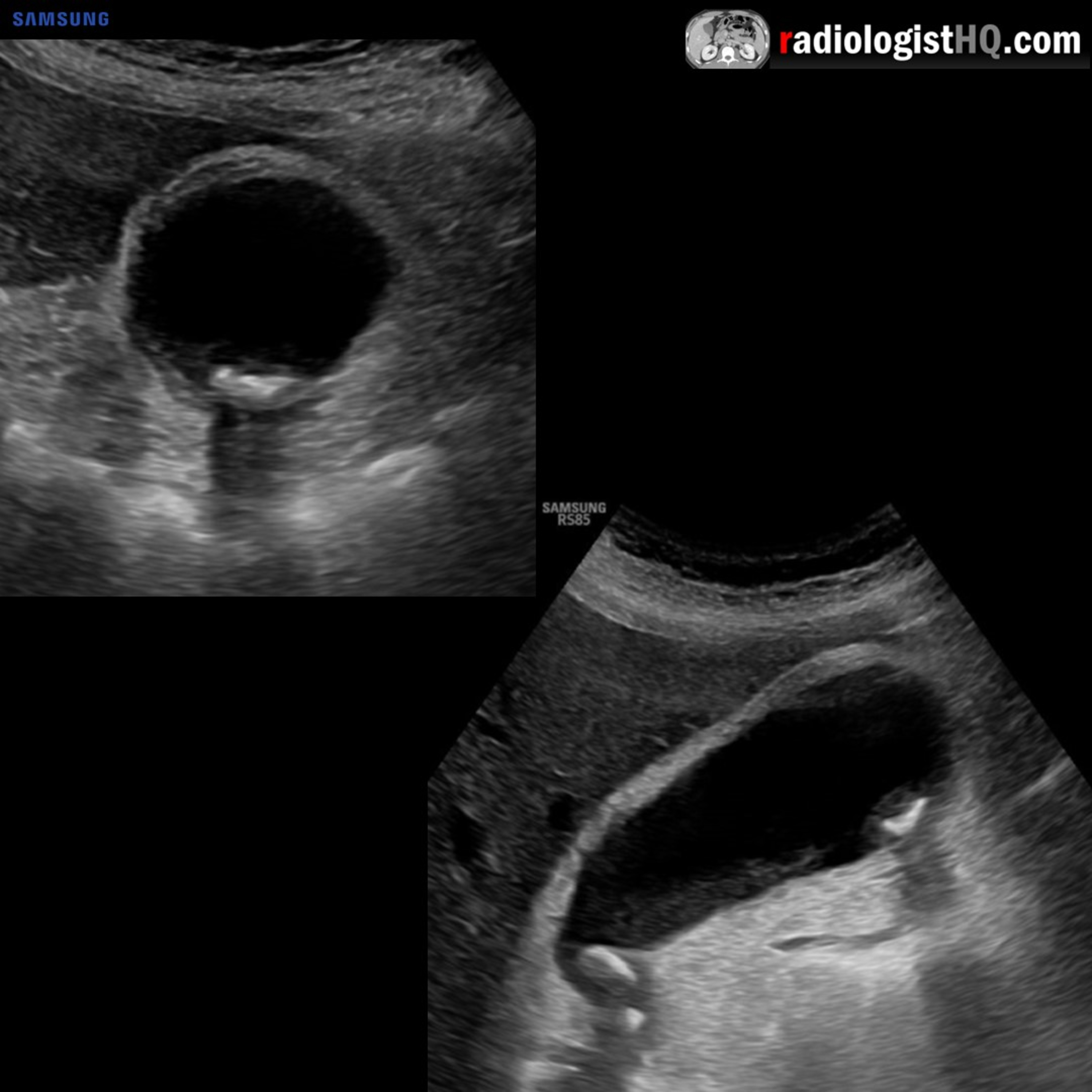
In this radiology lecture, we review the ultrasound appearance of acute cholecystitis, including gangrenous and emphysematous cholecystitis! Key teaching points include: Acute cholecystitis = Acute gallbladder inflammation. Most often (95%) caused by an impacted, obstructing gallstone in the cystic duct or gallbladder neck = Acute calculous cholecystitis. Clinically presents as persistent RUQ pain that may radiate to right shoulder, often with N/V and fever. Ultrasound findings of uncomplicated acute cholecystitis: Gallstones, sonographic Murphy sign, gallbladder wall thickening (greater than 3 mm) and edema, gallbladder distention (greater than 4 cm short axis), and pericholecystic fluid. Sonographic Murphy sign = Maximal abdominal tenderness from transducer pressure over gallbladder. PPV of gallstones and a positive sonographic Murphy sign = 92%. Pericholecystic fluid occurs in less than 20% of patients with acute cholecystitis, usually seen in more advanced cases. Gangrenous cholecystitis = Most common complication of acute cholecystitis. Ischemia with necrosis of gallbladder wall. Increased mortality compared to uncomplicated acute cholecystitis. Ultrasound findings of gangrenous cholecystitis: Wall disruption, ulceration, mucosal irregularity, and/or focal bulge, sloughed mucosal membranes, pericholecystic fluid, less likely to have positive Murphy sign, and increased risk of perforation (usually at fundus). Emphysematous cholecystitis = Gallbladder wall necrosis with gas formation in wall and/or lumen. More common in elderly men with underlying diabetes. Higher risk of perforation, rapid progression, and increased mortality compared to uncomplicated acute cholecystitis. Emergent surgical intervention typically required. Ultrasound findings of emphysematous cholecystitis: Bright reflectors from nondependent portions of gallbladder wall, dirty posterior acoustic shadowing, and ring-down artifact. CT can confirm if necessary. To learn more about the Samsung RS85 Prestige ultrasound system, please visit: ⁠⁠⁠https://www.bostonimaging.com/rs85-prestige-ultrasound-system-4⁠⁠⁠ Click the ⁠⁠⁠YouTube Community tab⁠⁠⁠ or follow on social media for bonus teaching material posted throughout the week! Website: ⁠⁠⁠https://radiologisthq.com/⁠⁠⁠ Instagram: ⁠⁠⁠https://www.instagram.com/radiologistHQ/⁠⁠⁠ Facebook: ⁠⁠⁠https://www.facebook.com/radiologistHeadQuarters/⁠⁠⁠ Twitter: ⁠⁠⁠https://twitter.com/radiologistHQ⁠⁠⁠ Reddit: ⁠⁠⁠https://www.reddit.com/user/radiologistHQ/

In this radiology lecture, we review the ultrasound appearance of acute cholecystitis, including gangrenous and emphysematous cholecystitis! Key teaching points include: Acute cholecystitis = Acute gallbladder inflammation. Most often (95%) caused by an impacted, obstructing gallstone in the cystic duct or gallbladder neck = Acute calculous cholecystitis. Clinically presents as persistent RUQ pain that may radiate to right shoulder, often with N/V and fever. Ultrasound findings of uncomplicated acute cholecystitis: Gallstones, sonographic Murphy sign, gallbladder wall thickening (greater than 3 mm) and edema, gallbladder distention (greater than 4 cm short axis), and pericholecystic fluid. Sonographic Murphy sign = Maximal abdominal tenderness from transducer pressure over gallbladder. PPV of gallstones and a positive sonographic Murphy sign = 92%. Pericholecystic fluid occurs in less than 20% of patients with acute cholecystitis, usually seen in more advanced cases. Gangrenous cholecystitis = Most common complication of acute cholecystitis. Ischemia with necrosis of gallbladder wall. Increased mortality compared to uncomplicated acute cholecystitis. Ultrasound findings of gangrenous cholecystitis: Wall disruption, ulceration, mucosal irregularity, and/or focal bulge, sloughed mucosal membranes, pericholecystic fluid, less likely to have positive Murphy sign, and increased risk of perforation (usually at fundus). Emphysematous cholecystitis = Gallbladder wall necrosis with gas formation in wall and/or lumen. More common in elderly men with underlying diabetes. Higher risk of perforation, rapid progression, and increased mortality compared to uncomplicated acute cholecystitis. Emergent surgical intervention typically required. Ultrasound findings of emphysematous cholecystitis: Bright reflectors from nondependent portions of gallbladder wall, dirty posterior acoustic shadowing, and ring-down artifact. CT can confirm if necessary. To learn more about the Samsung RS85 Prestige ultrasound system, please visit: ⁠⁠⁠https://www.bostonimaging.com/rs85-prestige-ultrasound-system-4⁠⁠⁠ Click the ⁠⁠⁠YouTube Community tab⁠⁠⁠ or follow on social media for bonus teaching material posted throughout the week! Website: ⁠⁠⁠https://radiologisthq.com/⁠⁠⁠ Instagram: ⁠⁠⁠https://www.instagram.com/radiologistHQ/⁠⁠⁠ Facebook: ⁠⁠⁠https://www.facebook.com/radiologistHeadQuarters/⁠⁠⁠ Twitter: ⁠⁠⁠https://twitter.com/radiologistHQ⁠⁠⁠ Reddit: ⁠⁠⁠https://www.reddit.com/user/radiologistHQ/

NOW PLAYING

Ultrasound of Acute Cholecystitis

0:00 10:57

No transcript for this episode yet

We transcribe on demand. Request one and we'll notify you when it's ready — usually under 10 minutes.

Frequently Asked Questions

How long is this episode of Radiology Lectures | Radquarters?

This episode is 10 minutes long.

When was this Radiology Lectures | Radquarters episode published?

This episode was published on July 27, 2023.

What is this episode about?

In this radiology lecture, we review the ultrasound appearance of acute cholecystitis, including gangrenous and emphysematous cholecystitis! Key teaching points include: Acute cholecystitis = Acute gallbladder inflammation. Most often (95%)...

Can I download this Radiology Lectures | Radquarters episode?

Yes, you can download this episode by clicking the download button on the episode player, or subscribe to the podcast in your preferred podcast app for automatic downloads.
URL copied to clipboard!