Ultrasound of Gout episode artwork

EPISODE · Oct 30, 2025 · 8 MIN

Ultrasound of Gout

from Radiology Lectures | Radquarters · host Daniel J. Kowal, MD

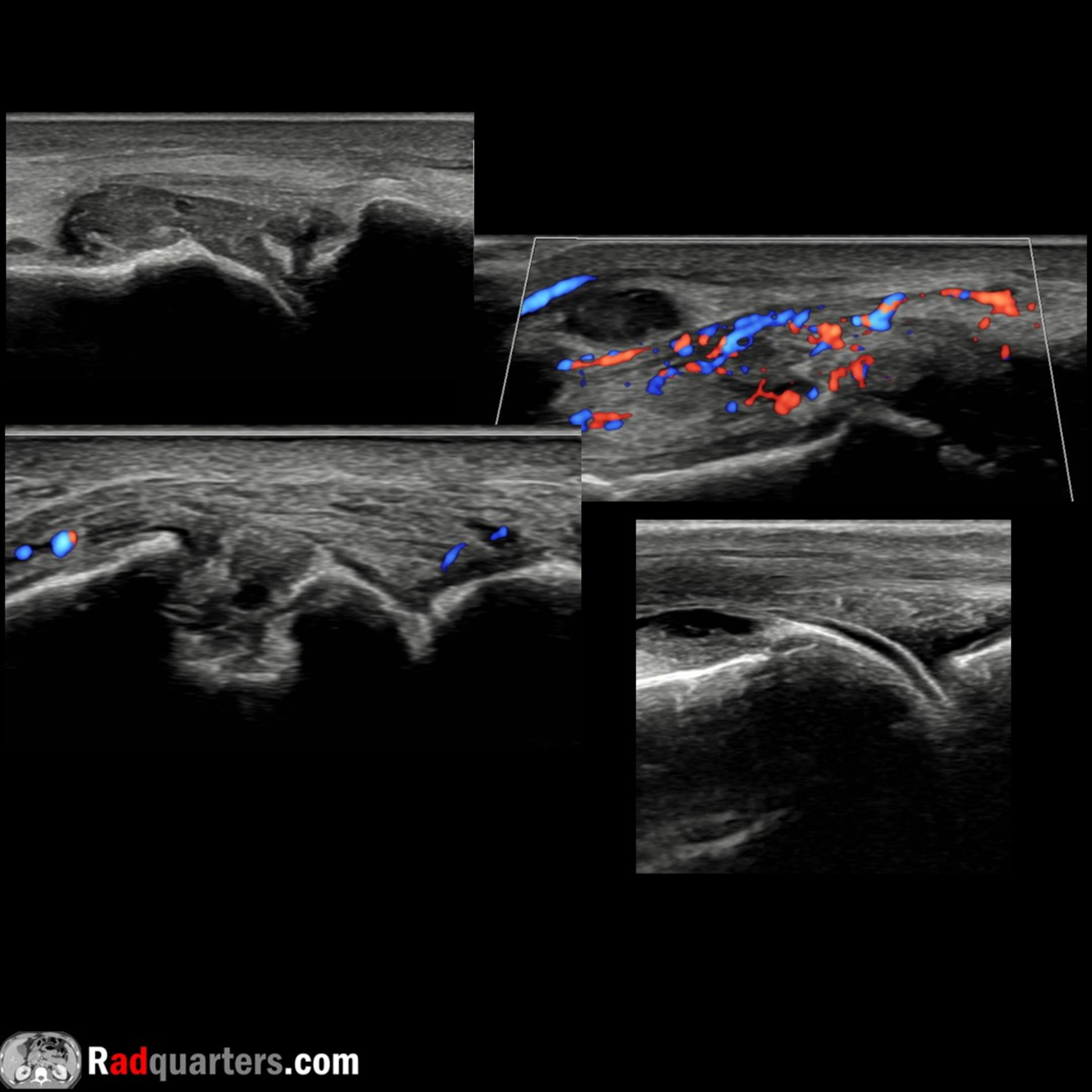
In this radiology lecture, we review the ultrasound appearance of gout!Key teaching points include:Gout is a crystal arthropathy due to monosodium urate crystal deposition in and around jointsMost common in males over age 40Risk factors: Metabolic (hyperuricemia, obesity, diabetes, hypertension), renal (chronic kidney disease), dietary (high purine foods, sugary drinks, alcohol), and genetic (family history)Typically presents as acute monoarthritis with a red, inflamed, swollen joint. First metatarsophalangeal joint most common site of involvement (podagra)Can progress to asymmetric polyarticular disease and chronic tophaceous goutUltrasound findings include: Joint effusion +/- hyperechoic foci (crystals/microtophi), synovial hypertrophy, and erosions typically at medial aspect of distal first metatarsal Erosions are juxtaarticular in distribution with overhanging edges yielding a “punched-out” appearanceGouty tophus: Amorphous, echogenic area containing internal hyperechoic foci surrounded by an anechoic inflammatory halo. May have associated cortical erosionsTophi may involve tendons, tendon sheaths, and bursae. Other common sites include olecranon region (elbow), patellar and popliteal tendons (knee)Double contour sign, AKA urate icing: Hyperechoic monosodium urate crystals coating hyaline cartilage surface. Disappears when serum urate levels drop below 6 mg/dL*Distinct from chondrocalcinosis seen in calcium pyrophosphate deposition disease which will have echogenic crystals within cartilage as opposed to on surfaceReferences: 1) *Thiele RG, Schlesinger N. Ultrasonography shows disappearance of monosodium urate crystal deposition on hyaline cartilage after sustained normouricemia is achieved. Rheumatol Int. 2010;30(4):495–5032) Jacobson JA. Fundamentals of Musculoskeletal Ultrasound. 3rd ed. Elsevier; 2018To learn more about the Samsung RS85 Prestige ultrasound system, please visit: ⁠⁠⁠⁠⁠⁠⁠⁠⁠⁠⁠⁠⁠⁠⁠https://www.bostonimaging.com/rs85-prestige-ultrasound-system-4⁠⁠⁠⁠⁠⁠⁠⁠⁠⁠⁠⁠⁠⁠⁠⁠Click the ⁠YouTube posts tab⁠ or follow on social media for bonus teaching material posted throughout the week!Website: ⁠⁠⁠⁠⁠⁠⁠⁠⁠⁠⁠⁠⁠⁠⁠⁠https://radquarters.com/⁠⁠⁠⁠⁠⁠⁠⁠⁠⁠⁠⁠⁠⁠⁠⁠Instagram: ⁠⁠⁠⁠⁠⁠⁠⁠⁠⁠⁠⁠⁠⁠⁠⁠https://www.instagram.com/radquarters/⁠⁠⁠⁠⁠⁠⁠⁠⁠⁠⁠⁠⁠⁠⁠⁠Facebook: ⁠⁠⁠⁠⁠⁠⁠⁠⁠⁠⁠⁠⁠⁠⁠⁠https://www.facebook.com/radquarters/⁠⁠⁠⁠⁠⁠⁠⁠⁠⁠⁠⁠⁠⁠⁠⁠X (Twitter): ⁠⁠⁠⁠⁠⁠⁠⁠⁠⁠⁠⁠⁠⁠⁠⁠https://twitter.com/radquarters⁠⁠⁠⁠⁠⁠⁠⁠⁠⁠⁠⁠⁠⁠⁠⁠Reddit: ⁠⁠⁠⁠⁠⁠⁠⁠⁠⁠⁠⁠⁠⁠⁠⁠⁠https://www.reddit.com/user/radiologistHQ/⁠⁠⁠⁠⁠This video is for informational purposes only. It does not replace the advice or counsel of a doctor or health care professional.

In this radiology lecture, we review the ultrasound appearance of gout!Key teaching points include:Gout is a crystal arthropathy due to monosodium urate crystal deposition in and around jointsMost common in males over age 40Risk factors: Metabolic (hyperuricemia, obesity, diabetes, hypertension), renal (chronic kidney disease), dietary (high purine foods, sugary drinks, alcohol), and genetic (family history)Typically presents as acute monoarthritis with a red, inflamed, swollen joint. First metatarsophalangeal joint most common site of involvement (podagra)Can progress to asymmetric polyarticular disease and chronic tophaceous goutUltrasound findings include: Joint effusion +/- hyperechoic foci (crystals/microtophi), synovial hypertrophy, and erosions typically at medial aspect of distal first metatarsal Erosions are juxtaarticular in distribution with overhanging edges yielding a “punched-out” appearanceGouty tophus: Amorphous, echogenic area containing internal hyperechoic foci surrounded by an anechoic inflammatory halo. May have associated cortical erosionsTophi may involve tendons, tendon sheaths, and bursae. Other common sites include olecranon region (elbow), patellar and popliteal tendons (knee)Double contour sign, AKA urate icing: Hyperechoic monosodium urate crystals coating hyaline cartilage surface. Disappears when serum urate levels drop below 6 mg/dL*Distinct from chondrocalcinosis seen in calcium pyrophosphate deposition disease which will have echogenic crystals within cartilage as opposed to on surfaceReferences: 1) *Thiele RG, Schlesinger N. Ultrasonography shows disappearance of monosodium urate crystal deposition on hyaline cartilage after sustained normouricemia is achieved. Rheumatol Int. 2010;30(4):495–5032) Jacobson JA. Fundamentals of Musculoskeletal Ultrasound. 3rd ed. Elsevier; 2018To learn more about the Samsung RS85 Prestige ultrasound system, please visit: ⁠⁠⁠⁠⁠⁠⁠⁠⁠⁠⁠⁠⁠⁠⁠https://www.bostonimaging.com/rs85-prestige-ultrasound-system-4⁠⁠⁠⁠⁠⁠⁠⁠⁠⁠⁠⁠⁠⁠⁠⁠Click the ⁠YouTube posts tab⁠ or follow on social media for bonus teaching material posted throughout the week!Website: ⁠⁠⁠⁠⁠⁠⁠⁠⁠⁠⁠⁠⁠⁠⁠⁠https://radquarters.com/⁠⁠⁠⁠⁠⁠⁠⁠⁠⁠⁠⁠⁠⁠⁠⁠Instagram: ⁠⁠⁠⁠⁠⁠⁠⁠⁠⁠⁠⁠⁠⁠⁠⁠https://www.instagram.com/radquarters/⁠⁠⁠⁠⁠⁠⁠⁠⁠⁠⁠⁠⁠⁠⁠⁠Facebook: ⁠⁠⁠⁠⁠⁠⁠⁠⁠⁠⁠⁠⁠⁠⁠⁠https://www.facebook.com/radquarters/⁠⁠⁠⁠⁠⁠⁠⁠⁠⁠⁠⁠⁠⁠⁠⁠X (Twitter): ⁠⁠⁠⁠⁠⁠⁠⁠⁠⁠⁠⁠⁠⁠⁠⁠https://twitter.com/radquarters⁠⁠⁠⁠⁠⁠⁠⁠⁠⁠⁠⁠⁠⁠⁠⁠Reddit: ⁠⁠⁠⁠⁠⁠⁠⁠⁠⁠⁠⁠⁠⁠⁠⁠⁠https://www.reddit.com/user/radiologistHQ/⁠⁠⁠⁠⁠This video is for informational purposes only. It does not replace the advice or counsel of a doctor or health care professional.

NOW PLAYING

Ultrasound of Gout

0:00 8:26

No transcript for this episode yet

We transcribe on demand. Request one and we'll notify you when it's ready — usually under 10 minutes.

Frequently Asked Questions

How long is this episode of Radiology Lectures | Radquarters?

This episode is 8 minutes long.

When was this Radiology Lectures | Radquarters episode published?

This episode was published on October 30, 2025.

What is this episode about?

In this radiology lecture, we review the ultrasound appearance of gout!Key teaching points include:Gout is a crystal arthropathy due to monosodium urate crystal deposition in and around jointsMost common in males over age 40Risk factors: Metabolic...

Can I download this Radiology Lectures | Radquarters episode?

Yes, you can download this episode by clicking the download button on the episode player, or subscribe to the podcast in your preferred podcast app for automatic downloads.
URL copied to clipboard!