
EPISODE · Dec 15, 2022 · 8 MIN
Ultrasound of Hashimoto’s Thyroiditis
from Radiology Lectures | Radquarters · host Daniel J. Kowal, MD
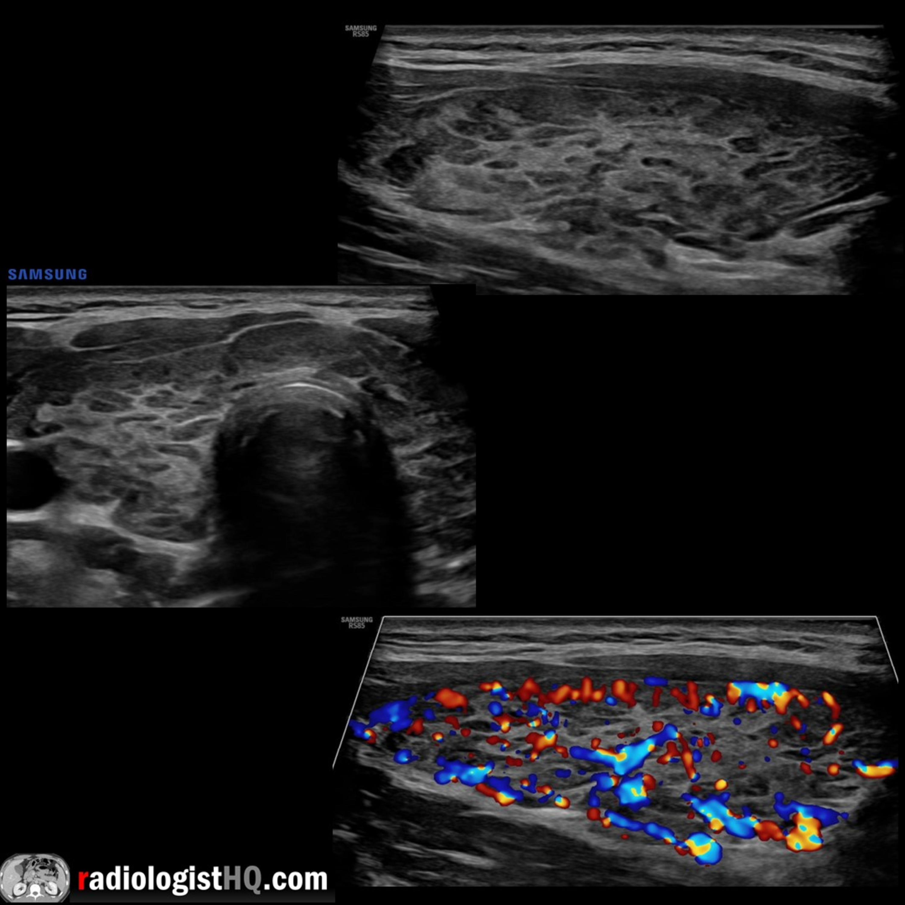
In this radiology lecture, we review the ultrasound appearance of Hashimoto’s thyroiditis with three unique cases! Key teaching points include: Normal thyroid gland isthmus measures less than 0.4 cm, transverse and AP lobe diameters measure less than 2 cm. Hashimoto’s thyroiditis is an autoimmune thyroiditis caused by antibodies to thyroid proteins. Most common in middle-aged females. May coexist with other autoimmune disorders: Lupus, rheumatoid arthritis. AKA chronic autoimmune lymphocytic thyroiditis: Gland is infiltrated with lymphocytes and plasma cells, fibrotic reaction replaces normal parenchyma. Leads to hypothyroidism = Most common cause in USA. Increased risk of thyroid cancer, including thyroid lymphoma. On ultrasound, gland is normal-sized or enlarged in initial phase with heterogeneously hypoechoic parenchymal echotexture. May have hypoechoic micronodules (1-6 mm) yielding a “pseudonodular” or “giraffe” pattern = High positive predictive value. Can also present with thin echogenic fibrous strands, lobulated contour, and geographic hypoechogenicity without discrete nodules. Gland may be atrophic in chronic cases. Variable color Doppler flow, may be hypervascular. Reactive, morphologically-normal neck nodes may be present. Can be difficult to differentiate from other forms of thyroiditis on ultrasound. Laboratory/serologic diagnosis: Thyroid function tests (TSH, free T4 test), thyroid peroxidase (TPO) antibodies present in most (95%) patients, and antithyroglobulin antibodies. Treatment: Thyroid hormone replacement if hypothyroid. To learn more about the Samsung RS85 Prestige ultrasound system, please visit: https://www.bostonimaging.com/rs85-prestige-ultrasound-system-4 Click the YouTube Community tab or follow on social media for bonus teaching material posted throughout the week! Website: https://radiologisthq.com/ Instagram: https://www.instagram.com/radiologistHQ/ Facebook: https://www.facebook.com/radiologistHeadQuarters/ Twitter: https://twitter.com/radiologistHQ Reddit: https://www.reddit.com/user/radiologistHQ/
Embed this episode
Ready to play
Ultrasound of Hashimoto’s Thyroiditis
No transcript for this episode yet
Similar Episodes
No similar episodes found.