
EPISODE · Apr 4, 2024 · 6 MIN
Ultrasound of Parathyroid Adenoma
from Radiology Lectures | Radquarters · host Daniel J. Kowal, MD
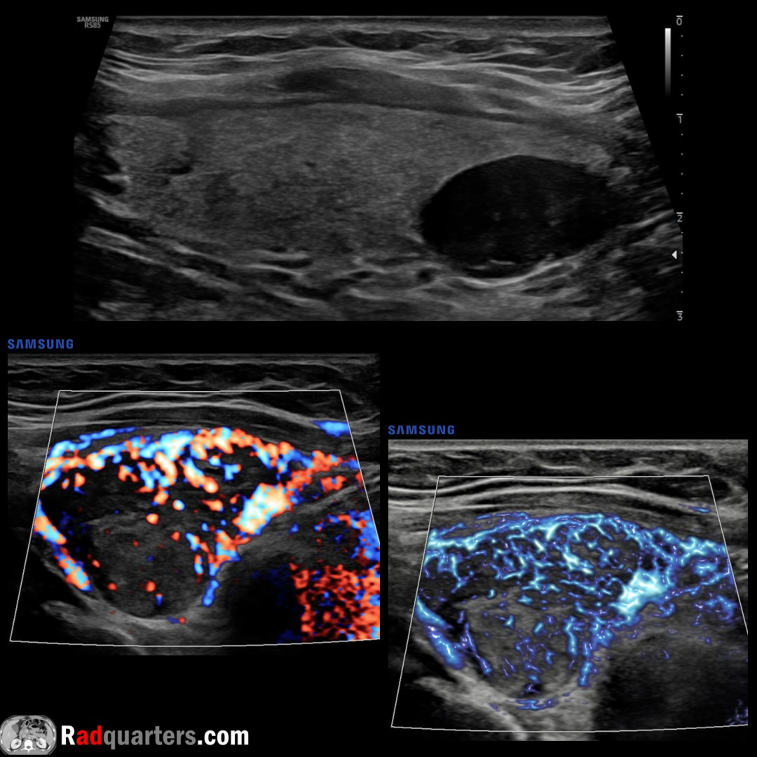
In this radiology lecture, we review the ultrasound appearance of parathyroid adenoma! Key teaching points include: Benign tumor of the parathyroid glands Most common cause of primary hyperparathyroidism: Elevated serum calcium and parathyroid hormone (PTH) levels Ultrasound: Solid, homogeneous and very hypoechoic. Oval or bean-shaped, long axis oriented craniocaudal. Hypervascular. Majority posterior and inferior to thyroid. Hyperechoic line often separates adenoma from adjacent thyroid. Atypical features: Cystic degeneration, calcification. Tc-99m sestamibi: Radiotracer uptake persisting on delayed 2-hour images. Taken up by both thyroid and parathyroid tissue, but washes out more rapidly from thyroid. Greater than 90% predictive value for preoperative localization of parathyroid adenoma. SPECT aids with anatomic localization Ectopic locations in up to 5%: Lower neck, mediastinum, retrotracheal/retroesophageal, carotid sheath and intrathyroidal (typically more homogeneous than thyroid nodules and have a linear interface with gland) Larger adenomas can be multilobulated “Polar vessel” sign: Enlarged feeding artery or draining vein terminating at parathyroid adenoma To learn more about the Samsung RS85 Prestige ultrasound system, please visit: https://www.bostonimaging.com/rs85-prestige-ultrasound-system-4 Click the YouTube Community tab or follow on social media for bonus teaching material posted throughout the week! Website: https://radquarters.com/ Instagram: https://www.instagram.com/radquarters/ Facebook: https://www.facebook.com/radquarters/ X (Twitter): https://twitter.com/radquarters Reddit: https://www.reddit.com/user/radiologistHQ/
Embed this episode
Ready to play
Ultrasound of Parathyroid Adenoma
No transcript for this episode yet
Similar Episodes
No similar episodes found.