
EPISODE · Feb 8, 2024 · 8 MIN
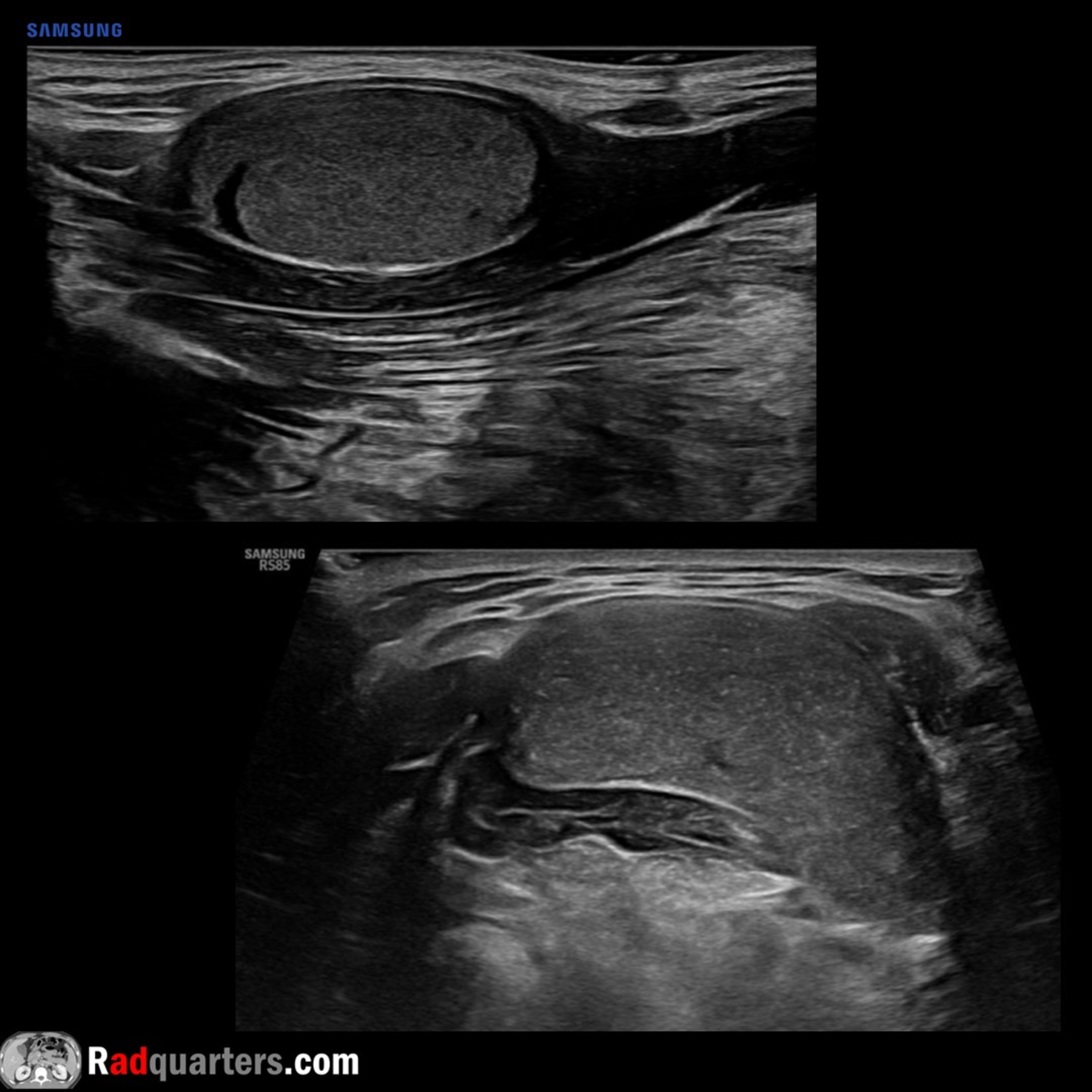
Ultrasound of Sublingual Dermoid Cyst
from Radiology Lectures | Radquarters · host Daniel J. Kowal, MD
In this radiology lecture, we review the ultrasound appearance of sublingual dermoid cyst and explain floor of mouth anatomy! Key teaching points include: The floor of the mouth is a horseshoe-shaped area beneath tongue and in between sides of mandible, inferiorly bounded by mylohyoid muscle, and containing sublingual space (SLS) SLS medial border: Midline genioglossus/geniohyoid muscle complex; SLS inferolateral border: Mylohyoid muscle Anterior margin of hyoglossus muscle projects into posterior SLS Sublingual dermoid cyst is a rare, benign cyst with squamous epithelial lining and contains skin appendages Dermoid and epidermoid cysts are in same family, terminology often used interchangeably, although epidermoid cysts less common and tend to contain fluid contents only Dermoid cyst mean age of presentation late teens to twenties, average age 30 Presents as a slowly enlarging neck mass, may cause dysphagia Often round or oval in shape and homogeneously hypoechoic with punctate echogenic foci May have pathognomonic “sack of marbles” appearance Relationship to mylohyoid is key for surgical planning: Intraoral resection for sublingual (above mylohyoid) location, extraoral approach for submental/submandibular (below mylohyoid) location Most cysts are midline DDx: Suprahyoid thyroglossal duct cyst, ranula (simple and diving), abscess and lymphangioma To learn more about the Samsung RS85 Prestige ultrasound system, please visit: https://www.bostonimaging.com/rs85-prestige-ultrasound-system-4 Click the YouTube Community tab or follow on social media for bonus teaching material posted throughout the week! Website: https://radquarters.com/ Instagram: https://www.instagram.com/radquarters/ Facebook: https://www.facebook.com/radquarters/ X (Twitter): https://twitter.com/radquarters Reddit: https://www.reddit.com/user/radiologistHQ/
Embed this episode
Ready to play
Ultrasound of Sublingual Dermoid Cyst
No transcript for this episode yet
Similar Episodes
No similar episodes found.