
EPISODE · Jul 14, 2024 · 9 MIN
Ultrasound of Tennis Leg
from Radiology Lectures | Radquarters · host Daniel J. Kowal, MD
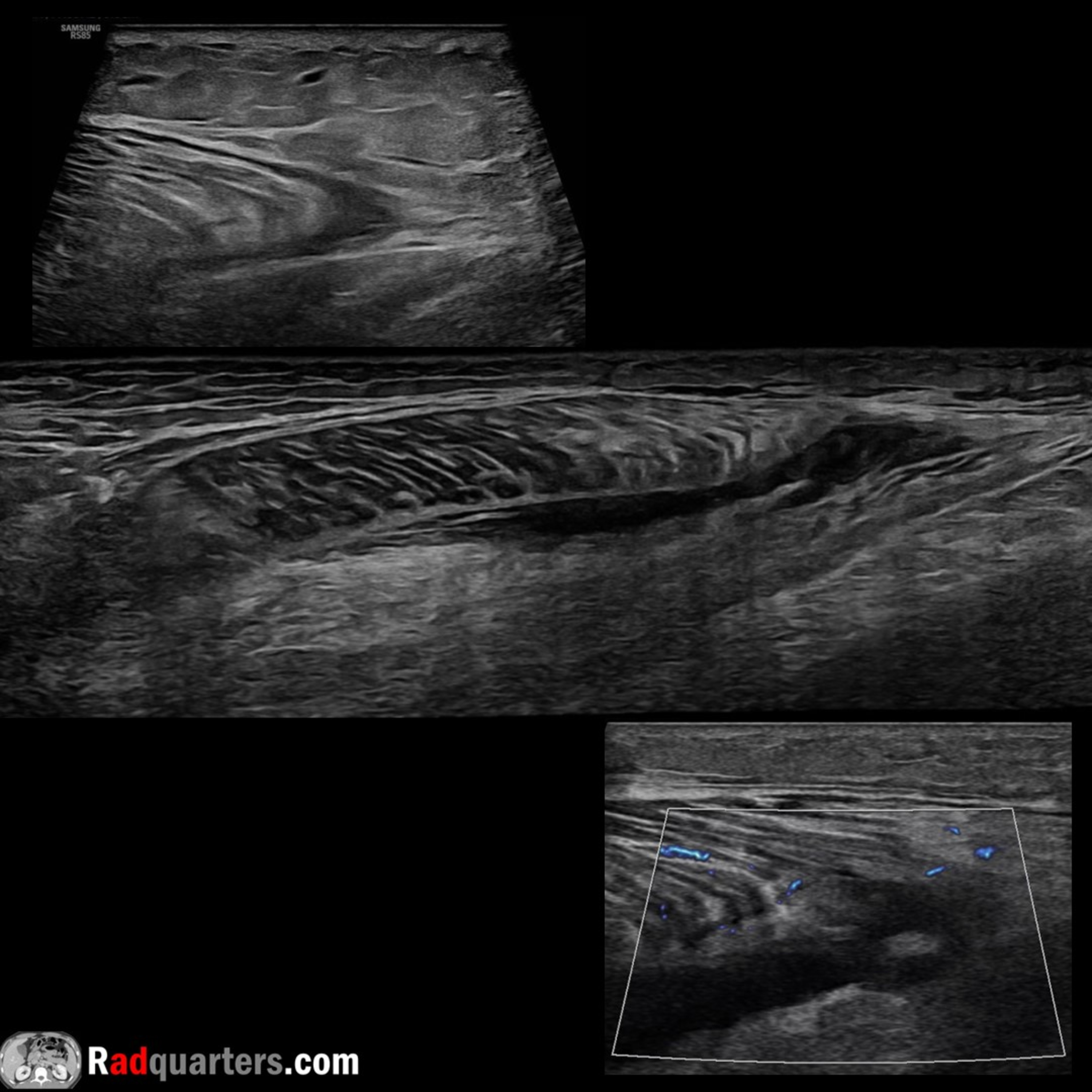
In this radiology lecture, we review the ultrasound appearance of tennis leg, including medial gastrocnemius and plantaris injury! Key teaching points include: Tennis leg = Injury to muscles of the calf. Tear of myotendinous junction of medial head of gastrocnemius, rupture of plantaris tendon (less common), in isolation or together Classically described in tennis players, but can occur in various athletic activities (running, skiing) with extension of knee and forced dorsiflexion of ankle. Typically seen in middle-aged, active individuals Clinical: Sudden sharp calf pain with associated popping/snapping sensation followed by tenderness and swelling Gastrocnemius & soleus are pennate muscles. Fascicles attach obliquely to a tendon = Aponeuroses with long length of musculotendinous junction. Feathers converging on a single point Triceps surae muscle = Two headed gastrocnemius, soleus and plantaris. Distal continuation of the gastrocnemius and soleus forms the Achilles tendon Distal medial head of gastrocnemius where tapers over soleus = One of most commonly injured calf structures Medial gastrocnemius tear appears as disrupted tendon fibers at aponeurosis with anechoic/hypoechoic fluid or hemorrhage +/- muscle retraction May see retracted muscle fascicles. Hematoma can dissect between and extends into medial gastrocnemius and soleus muscles Tx: Conservative (self-limiting). Surgical fasciotomy if compartment syndrome Plantaris muscle arises from the posterosuperior aspect of lateral femoral condyle near lateral head origin of gastrocnemius muscle. Medially crosses posterior knee joint in oblique fashion Plantaris continues into calf as a long, thin tendon traveling between medial head of gastrocnemius and soleus muscles. Courses distally at medial aspect of Achilles tendon, usually inserts onto calcaneus. Plantaris is absent in up to 20% Plantaris injury/rupture less common than medial head gastrocnemius tear and typically more proximal in calf (at myotendinous junction) To learn more about the Samsung RS85 Prestige ultrasound system, please visit: https://www.bostonimaging.com/rs85-prestige-ultrasound-system-4 Click the YouTube Community tab or follow on social media for bonus teaching material posted throughout the week! Website: https://radquarters.com/ Instagram: https://www.instagram.com/radquarters/ Facebook: https://www.facebook.com/radquarters/ X (Twitter): https://twitter.com/radquarters Reddit: https://www.reddit.com/user/radiologistHQ/
Embed this episode
Ready to play
Ultrasound of Tennis Leg
No transcript for this episode yet
Similar Episodes
No similar episodes found.