
EPISODE · Aug 31, 2022 · 7 MIN
Ultrasound of Varicocele
from Radiology Lectures | Radquarters · host Daniel J. Kowal, MD
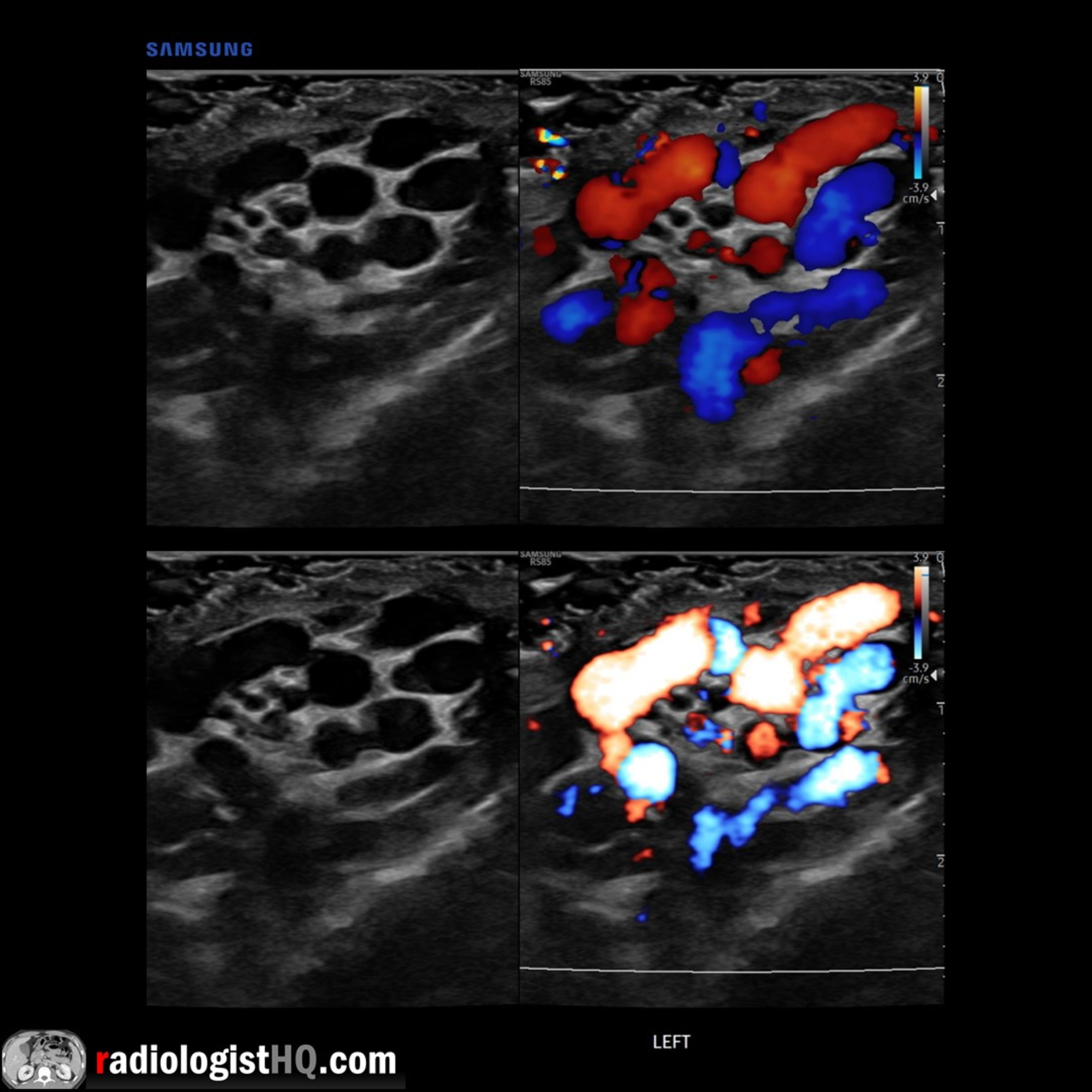
In this radiology lecture, we review the ultrasound appearance of scrotal varicocele with three unique cases. Key teaching points include: Varicocele is abnormal dilatation of pampiniform venous plexus = Peritesticular veins. Seen in up to 15% of adult and adolescent males. Caused by incompetent or absent testicular vein valves. Upper limit of normal for scrotal vein caliber = 2 mm, varicocele when greater than 2-3 mm. Flow in varicocele usually too slow to detect with color Doppler and is typically better seen with Valsalva or with standing position. 85% left sided, 15% bilateral: Left testicular vein drains into left renal vein at 90-degree angle, and superior mesenteric artery compresses left renal vein = Increased pressure and venous backflow. Right vein drains into IVC at acute angle. Symptoms: Scrotal mass, pain, infertility/subfertility. Low grade: Reflux only seen with Valsalva, inguinal canal/supratesticular location, vessels enlarged only in standing position. High grade: Reflux seen at rest, infratesticular location, vessels enlarged in supine position. Solitary right varicocele raises concern for compression of the right testicular vein from a retroperitoneal mass. Ultrasound of upper abdomen should be considered when an isolated right-sided varicocele or asymmetrically large right-sided varicocele found. However, most patients typically present with additional signs and symptoms of malignancy: “No patient in our cohort was found to have an unsuspected malignancy for which isolated right-sided varicocele was the only presenting sign.”* *Gleason A, Bishop K, Xi Y et al. Isolated Right-Sided Varicocele: Is Further Workup Necessary? AJR 2019; 212:802-807 To learn more about the Samsung RS85 Prestige ultrasound system, please visit: https://www.bostonimaging.com/rs85-prestige-ultrasound-system-4 Click the YouTube Community tab or follow on social media for bonus teaching material posted throughout the week! Website: https://radiologisthq.com/ Instagram: https://www.instagram.com/radiologistHQ/ Facebook: https://www.facebook.com/radiologistHeadQuarters/ Twitter: https://twitter.com/radiologistHQ Reddit: https://www.reddit.com/user/radiologistHQ/
Embed this episode
Ready to play
Ultrasound of Varicocele
No transcript for this episode yet
Similar Episodes
No similar episodes found.