EPISODE · Nov 25, 2023 · 4 MIN
“What are Differential Diagnoses for the Pyogenic Granuloma?” - Quick Review #74
from Dr. Gallagher's Podcast · host Brendan Gallagher, DDS
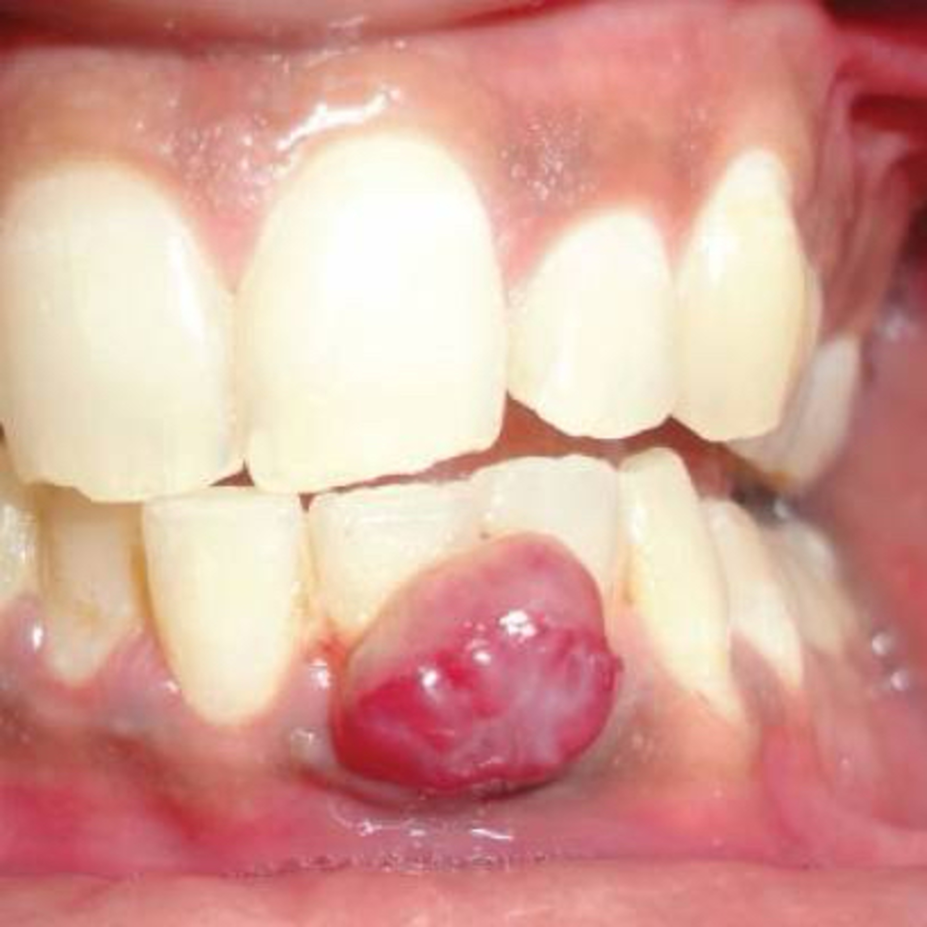
11.25.23 #pathology #oralpathology #doctorgallagher #oralsurgery #oralsurgeon #dentist #dentistry #dental Oral pyogenic granuloma is a relatively common benign mucosal lesion, often appearing as a red, soft, and painless nodule that may bleed easily. It’s important to distinguish it from other oral lesions that can present similarly: 1. Peripheral Giant Cell Granuloma (PGCG): •Defining Features: 1. Exclusively occurs on the gingiva, often in areas with dental irritation or trauma. 2. Presents as a red-purple nodule, which can be sessile or pedunculated. 3. Histologically, it contains multinucleated giant cells, hemorrhage, and hemosiderin deposits. •Similarities to Oral Pyogenic Granuloma: Both can present as red, vascular-appearing lesions on the gums. 2. Peripheral Ossifying Fibroma (POF): •Defining Features: 1. A fibrous growth that originates in the gingival interdental papilla, particularly in response to irritation. 2. Typically appears as a firm, nodular mass that can be pink to red. 3. Histologically, it contains fibrous tissue and can have areas of calcification or ossification. •Similarities: Both lesions are commonly found on the gingiva and can appear as a localized growth. 3. Squamous Cell Carcinoma: •Defining Features: 1. An invasive malignant tumor with potential for significant local destruction and metastasis. 2. Appears as a non-healing ulcer or a mass, which can be white, red, or mixed in color. 3. Histopathology reveals atypical squamous cells invading deeper tissue layers. •Similarities: Both can present as ulcerative lesions in the oral cavity. 4. Kaposi’s Sarcoma: •Defining Features: 1. Associated with Human Herpesvirus 8 (HHV-8), more common in immunocompromised patients. 2. Presents as red, purple, or brown macules, plaques, or nodules. 3. Histopathology shows spindle-shaped cells with vascular proliferation. 5. Fibroma: •Defining Features: 1. A common benign tumor composed of fibrous or connective tissue. 2. Typically presents as a small, smooth, pink, firm nodule. 3. Often occurs in response to trauma or irritation. 6. Hemangioma: •Defining Features: 1. A benign tumor of blood vessels. 2. Presents as a red to bluish, soft, compressible mass. 3. Often present at birth or appears in early childhood. References: 1. Jafarzadeh, H., Sanatkhani, M., & Mohtasham, N. (n.d.). Oral pyogenic granuloma: a review. PubMed. Retrieved from https://lnkd.in/eabFuPmb 2. Rowe, L. (1958). Granuloma Pyogenicum: Differential Diagnosis. JAMA Dermatology, 78(3), 341-347. doi:10.1001/archderm.1958.01560090055013. 3. Franco-Barrera, M. J., Zavala-Cerna, M. G., Fernández-Tamayo, R., Vivanco-Pérez, I., Fernández-Tamayo, N. M., Torres-Bugarín, O. (n.d.). An update on peripheral ossifying fibroma: case report and literature review. PubMed. Retrieved from https://lnkd.in/eabFuPmb 4. ChatGPT. 2023.
What this episode covers
11.25.23 #pathology #oralpathology #doctorgallagher #oralsurgery #oralsurgeon #dentist #dentistry #dental Oral pyogenic granuloma is a relatively common benign mucosal lesion, often appearing as a red, soft, and painless nodule that may bleed easily. It’s important to distinguish it from other oral lesions that can present similarly: 1. Peripheral Giant Cell Granuloma (PGCG): •Defining Features: 1. Exclusively occurs on the gingiva, often in areas with dental irritation or trauma. 2. Presents as a red-purple nodule, which can be sessile or pedunculated. 3. Histologically, it contains multinucleated giant cells, hemorrhage, and hemosiderin deposits. •Similarities to Oral Pyogenic Granuloma: Both can present as red, vascular-appearing lesions on the gums. 2. Peripheral Ossifying Fibroma (POF): •Defining Features: 1. A fibrous growth that originates in the gingival interdental papilla, particularly in response to irritation. 2. Typically appears as a firm, nodular mass that can be pink to red. 3. Histologically, it contains fibrous tissue and can have areas of calcification or ossification. •Similarities: Both lesions are commonly found on the gingiva and can appear as a localized growth. 3. Squamous Cell Carcinoma: •Defining Features: 1. An invasive malignant tumor with potential for significant local destruction and metastasis. 2. Appears as a non-healing ulcer or a mass, which can be white, red, or mixed in color. 3. Histopathology reveals atypical squamous cells invading deeper tissue layers. •Similarities: Both can present as ulcerative lesions in the oral cavity. 4. Kaposi’s Sarcoma: •Defining Features: 1. Associated with Human Herpesvirus 8 (HHV-8), more common in immunocompromised patients. 2. Presents as red, purple, or brown macules, plaques, or nodules. 3. Histopathology shows spindle-shaped cells with vascular proliferation. 5. Fibroma: •Defining Features: 1. A common benign tumor composed of fibrous or connective tissue. 2. Typically presents as a small, smooth, pink, firm nodule. 3. Often occurs in response to trauma or irritation. 6. Hemangioma: •Defining Features: 1. A benign tumor of blood vessels. 2. Presents as a red to bluish, soft, compressible mass. 3. Often present at birth or appears in early childhood. References: 1. Jafarzadeh, H., Sanatkhani, M., & Mohtasham, N. (n.d.). Oral pyogenic granuloma: a review. PubMed. Retrieved from https://lnkd.in/eabFuPmb 2. Rowe, L. (1958). Granuloma Pyogenicum: Differential Diagnosis. JAMA Dermatology, 78(3), 341-347. doi:10.1001/archderm.1958.01560090055013. 3. Franco-Barrera, M. J., Zavala-Cerna, M. G., Fernández-Tamayo, R., Vivanco-Pérez, I., Fernández-Tamayo, N. M., Torres-Bugarín, O. (n.d.). An update on peripheral ossifying fibroma: case report and literature review. PubMed. Retrieved from https://lnkd.in/eabFuPmb 4. ChatGPT. 2023.
NOW PLAYING
“What are Differential Diagnoses for the Pyogenic Granuloma?” - Quick Review #74
No transcript for this episode yet
Similar Episodes
Dec 5, 2025 ·50m
Oct 9, 2025 ·33m
Oct 3, 2025 ·40m
Sep 11, 2025 ·31m
Aug 27, 2025 ·39m
Aug 18, 2025 ·54m