EPISODE · Oct 18, 2024 · 4 MIN
📝 “What are Key Defining Features of the Odontogenic Keratocyst?”
from Dr. Gallagher's Podcast · host Brendan Gallagher, DDS
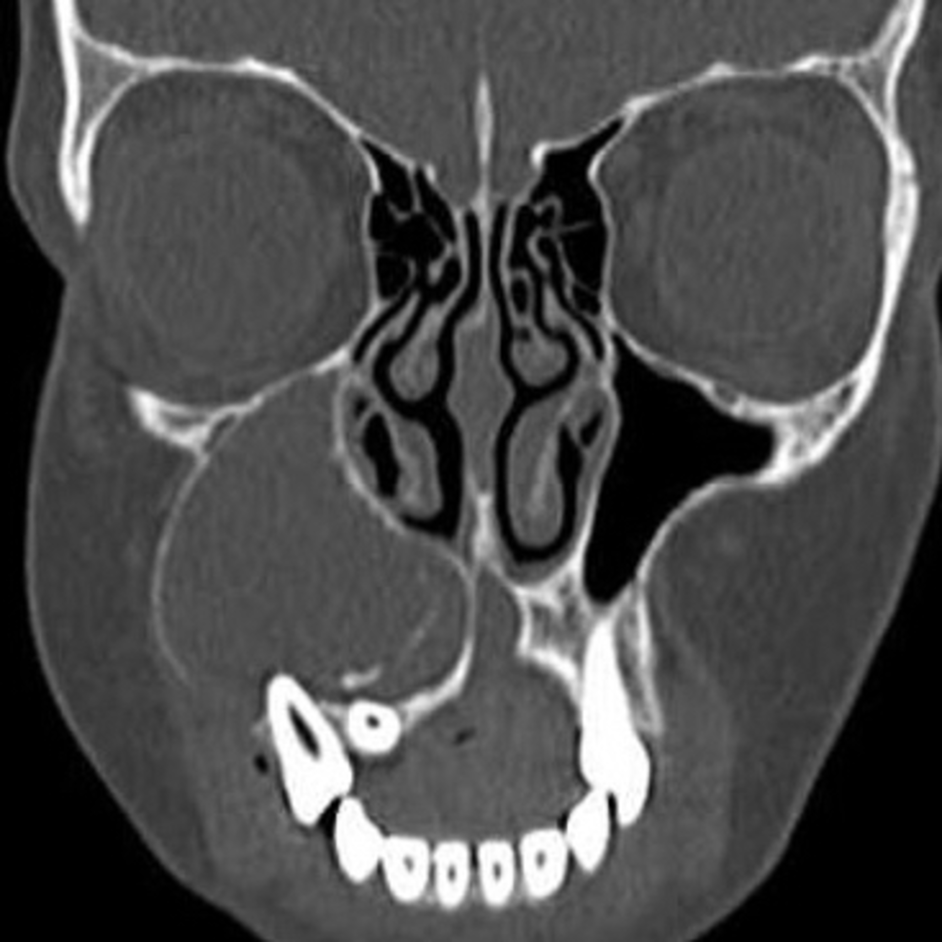
- 10.18.24Quick Review #247 - #pathology #oralpathology #doctorgallagher #oralsurgery #oralsurgeon #dentist #dentistry #dental Clinical Features:OKCs most commonly present between the second and third decades of life, with a slight male predilection. They can occur anywhere in the jaws but are most frequently found in the posterior mandible, particularly in the ramus and angle regions. Clinically, OKCs tend to grow along the anteroposterior axis of the bone, often causing minimal expansion until they reach significant size. Because of this growth pattern, patients may remain asymptomatic for a long time, and OKCs are frequently discovered incidentally during radiographic examinations. However, larger lesions may cause symptoms like pain, swelling, and, occasionally, drainage.Radiographic Features:Radiographically, OKCs can appear as well-defined radiolucencies, often with a smooth, corticated margin. The lesion may be unilocular or multilocular, and its size varies significantly. A unilocular appearance is more common in smaller cysts, whereas larger lesions tend to exhibit a multilocular pattern. In some cases, OKCs may be associated with an unerupted tooth, resembling a dentigerous cyst. Resorption of adjacent roots is rare, differentiating it from other odontogenic lesions like ameloblastomas.Histologic Features:Histologically, OKCs exhibit a distinctive lining composed of a uniform, thin layer of parakeratinized stratified squamous epithelium, which is typically about 6-8 cell layers thick. The epithelial surface shows a characteristic corrugated or wavy pattern. The basal cell layer is palisaded, meaning the nuclei are aligned in a vertical, fence-like fashion. The connective tissue wall is usually thin, and satellite cysts or daughter cysts are often present within it. Another notable histologic feature is the tendency of the cyst lining to separate from the connective tissue wall during surgical enucleation, which increases the risk of recurrence.Clinical Implications:OKCs are known for their high recurrence rate, which can range from 25% to 60% depending on the treatment method used. This is largely attributed to the difficulty of completely removing the cyst lining, the presence of satellite cysts, and its aggressive nature. Treatment typically involves enucleation and curettage, although more aggressive approaches like marsupialization or resection may be recommended in recurrent cases. Long-term follow-up is crucial due to the potential for recurrence.References:1. Niknejad, M. T. (n.d.). Maxillary odontogenic keratocyst. Radiopaedia. Retrieved October 17, 2024, from https://lnkd.in/eCt7Mh6f2. Neville, B. W., Damm, D. D., Allen, C. M., & Chi, A. C. (2015). Oral and Maxillofacial Pathology (4th ed.). Saunders. 3. ChstGPT. 2024.#podcast #podcasts #dentalpodcast #dentalpodcasts #doctorgallagherpodcast #doctorgallagherspodcast #doctor #dentistry #oralsurgery #dental #viral #dentalschool #dentalstudent #omfs #surgeon #doctorlife #dentistlife #residency #oralsurgeon #dentist #doctorgallagher
What this episode covers
- 10.18.24Quick Review #247 - #pathology #oralpathology #doctorgallagher #oralsurgery #oralsurgeon #dentist #dentistry #dental Clinical Features:OKCs most commonly present between the second and third decades of life, with a slight male predilection. They can occur anywhere in the jaws but are most frequently found in the posterior mandible, particularly in the ramus and angle regions. Clinically, OKCs tend to grow along the anteroposterior axis of the bone, often causing minimal expansion until they reach significant size. Because of this growth pattern, patients may remain asymptomatic for a long time, and OKCs are frequently discovered incidentally during radiographic examinations. However, larger lesions may cause symptoms like pain, swelling, and, occasionally, drainage.Radiographic Features:Radiographically, OKCs can appear as well-defined radiolucencies, often with a smooth, corticated margin. The lesion may be unilocular or multilocular, and its size varies significantly. A unilocular appearance is more common in smaller cysts, whereas larger lesions tend to exhibit a multilocular pattern. In some cases, OKCs may be associated with an unerupted tooth, resembling a dentigerous cyst. Resorption of adjacent roots is rare, differentiating it from other odontogenic lesions like ameloblastomas.Histologic Features:Histologically, OKCs exhibit a distinctive lining composed of a uniform, thin layer of parakeratinized stratified squamous epithelium, which is typically about 6-8 cell layers thick. The epithelial surface shows a characteristic corrugated or wavy pattern. The basal cell layer is palisaded, meaning the nuclei are aligned in a vertical, fence-like fashion. The connective tissue wall is usually thin, and satellite cysts or daughter cysts are often present within it. Another notable histologic feature is the tendency of the cyst lining to separate from the connective tissue wall during surgical enucleation, which increases the risk of recurrence.Clinical Implications:OKCs are known for their high recurrence rate, which can range from 25% to 60% depending on the treatment method used. This is largely attributed to the difficulty of completely removing the cyst lining, the presence of satellite cysts, and its aggressive nature. Treatment typically involves enucleation and curettage, although more aggressive approaches like marsupialization or resection may be recommended in recurrent cases. Long-term follow-up is crucial due to the potential for recurrence.References:1. Niknejad, M. T. (n.d.). Maxillary odontogenic keratocyst. Radiopaedia. Retrieved October 17, 2024, from https://lnkd.in/eCt7Mh6f2. Neville, B. W., Damm, D. D., Allen, C. M., & Chi, A. C. (2015). Oral and Maxillofacial Pathology (4th ed.). Saunders. 3. ChstGPT. 2024.#podcast #podcasts #dentalpodcast #dentalpodcasts #doctorgallagherpodcast #doctorgallagherspodcast #doctor #dentistry #oralsurgery #dental #viral #dentalschool #dentalstudent #omfs #surgeon #doctorlife #dentistlife #residency #oralsurgeon #dentist #doctorgallagher
NOW PLAYING
📝 “What are Key Defining Features of the Odontogenic Keratocyst?”
No transcript for this episode yet
Similar Episodes
Dec 5, 2025 ·50m
Oct 9, 2025 ·33m
Oct 3, 2025 ·40m
Sep 11, 2025 ·31m
Aug 27, 2025 ·39m
Aug 18, 2025 ·54m