EPISODE · Jul 12, 2024 · 4 MIN
“What are Key Differences Between Pemphigus & Pemphigoid?”
from Dr. Gallagher's Podcast · host Brendan Gallagher, DDS
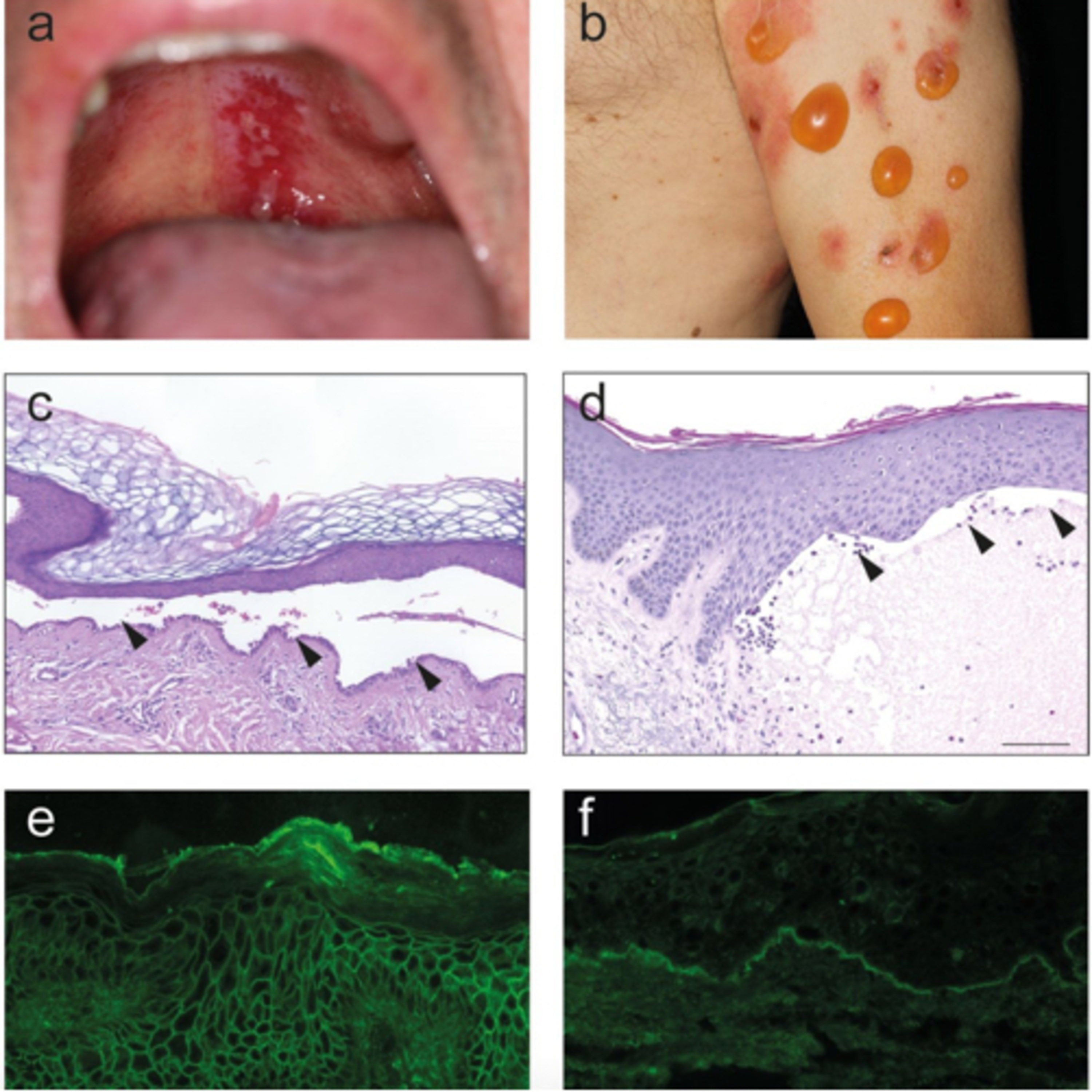
7.12.24 Quick Review #176 - #pathology #oralpathology #doctorgallagher #oralsurgery #oralsurgeon #dentist #dentistry #dental Pemphigus: 1. Pathogenesis: characterized by autoantibodies against desmogleins, proteins that form the intercellular junctions (desmosomes) between keratinocytes in the epidermis. This leads to a loss of cell-to-cell adhesion, resulting in intraepidermal blisters. 2. Types: • Pemphigus Vulgaris: The most common type, typically involves mucous membranes and skin. Blisters are flaccid and easily rupture, leaving painful erosions. • Pemphigus Foliaceus: Primarily affects the skin with superficial blisters, rarely involving mucous membranes. 3. Clinical Features: • Blisters and erosions on skin and mucous membranes. • Nikolsky sign positive (skin shears off easily when rubbed). 4. Histopathology: • Acantholysis (loss of intercellular connections) within the epidermis. • Intraepidermal blister formation. 5. Diagnosis: • Direct immunofluorescence showing intercellular deposition of IgG and C3 within the epidermis. • ELISA for desmoglein antibodies. Pemphigoid: 1. Pathogenesis: characterized by autoantibodies against components of the basement membrane zone, such as BP180 and BP230, leading to subepidermal blistering. 2. Types: • Bullous Pemphigoid: The most common type, typically affects elderly individuals and primarily involves the skin. • Mucous Membrane Pemphigoid: Also known as cicatricial pemphigoid, it predominantly affects mucous membranes and can lead to scarring. 3. Clinical Features: • Tense, less easily ruptured blisters on skin or mucous membranes. • Nikolsky sign usually negative. • Pruritus is common in bullous pemphigoid. 4. Histopathology: • Subepidermal blister formation. • Presence of inflammatory infiltrate, often with eosinophils, at the dermal-epidermal junction. 5. Diagnosis: • Direct immunofluorescence showing linear deposition of IgG and C3 at the basement membrane zone. • ELISA for BP180 and BP230 antibodies. References: 1. Bieber, K., Kridin, K., Emtenani, S., Boch, K., Schmidt, E., & Ludwig, R. J. (2021). Milestones in personalized medicine in pemphigus and pemphigoid. Frontiers in Immunology, 11, Article 591971. 2. Bolognia, J. L., Schaffer, J. V., & Cerroni, L. (2018). Dermatology (4th ed.). Elsevier. 3. James, W. D., Elston, D. M., Treat, J. R., & Rosenbach, M. A. (2020). Andrews’ Diseases of the Skin: Clinical Dermatology (13th ed.). Elsevier. 4. ChatGPT. 2024. - #podcast #podcasts #dentalpodcast #dentalpodcasts #doctorgallagherpodcast #doctorgallagherspodcast #doctor #dentistry #oralsurgery #dental #viral #dentalschool #dentalstudent #omfs #surgeon #doctorlife #dentistlife #residency #oralsurgeon #dentist #doctorgallagher
What this episode covers
7.12.24 Quick Review #176 - #pathology #oralpathology #doctorgallagher #oralsurgery #oralsurgeon #dentist #dentistry #dental Pemphigus: 1. Pathogenesis: characterized by autoantibodies against desmogleins, proteins that form the intercellular junctions (desmosomes) between keratinocytes in the epidermis. This leads to a loss of cell-to-cell adhesion, resulting in intraepidermal blisters. 2. Types: • Pemphigus Vulgaris: The most common type, typically involves mucous membranes and skin. Blisters are flaccid and easily rupture, leaving painful erosions. • Pemphigus Foliaceus: Primarily affects the skin with superficial blisters, rarely involving mucous membranes. 3. Clinical Features: • Blisters and erosions on skin and mucous membranes. • Nikolsky sign positive (skin shears off easily when rubbed). 4. Histopathology: • Acantholysis (loss of intercellular connections) within the epidermis. • Intraepidermal blister formation. 5. Diagnosis: • Direct immunofluorescence showing intercellular deposition of IgG and C3 within the epidermis. • ELISA for desmoglein antibodies. Pemphigoid: 1. Pathogenesis: characterized by autoantibodies against components of the basement membrane zone, such as BP180 and BP230, leading to subepidermal blistering. 2. Types: • Bullous Pemphigoid: The most common type, typically affects elderly individuals and primarily involves the skin. • Mucous Membrane Pemphigoid: Also known as cicatricial pemphigoid, it predominantly affects mucous membranes and can lead to scarring. 3. Clinical Features: • Tense, less easily ruptured blisters on skin or mucous membranes. • Nikolsky sign usually negative. • Pruritus is common in bullous pemphigoid. 4. Histopathology: • Subepidermal blister formation. • Presence of inflammatory infiltrate, often with eosinophils, at the dermal-epidermal junction. 5. Diagnosis: • Direct immunofluorescence showing linear deposition of IgG and C3 at the basement membrane zone. • ELISA for BP180 and BP230 antibodies. References: 1. Bieber, K., Kridin, K., Emtenani, S., Boch, K., Schmidt, E., & Ludwig, R. J. (2021). Milestones in personalized medicine in pemphigus and pemphigoid. Frontiers in Immunology, 11, Article 591971. 2. Bolognia, J. L., Schaffer, J. V., & Cerroni, L. (2018). Dermatology (4th ed.). Elsevier. 3. James, W. D., Elston, D. M., Treat, J. R., & Rosenbach, M. A. (2020). Andrews’ Diseases of the Skin: Clinical Dermatology (13th ed.). Elsevier. 4. ChatGPT. 2024. - #podcast #podcasts #dentalpodcast #dentalpodcasts #doctorgallagherpodcast #doctorgallagherspodcast #doctor #dentistry #oralsurgery #dental #viral #dentalschool #dentalstudent #omfs #surgeon #doctorlife #dentistlife #residency #oralsurgeon #dentist #doctorgallagher
NOW PLAYING
“What are Key Differences Between Pemphigus & Pemphigoid?”
No transcript for this episode yet
Similar Episodes
Dec 5, 2025 ·50m
Oct 9, 2025 ·33m
Oct 3, 2025 ·40m
Sep 11, 2025 ·31m
Aug 27, 2025 ·39m
Aug 18, 2025 ·54m