EPISODE · Dec 23, 2023 · 5 MIN
“What are the 3 LeFort Fracture Types?”
from Dr. Gallagher's Podcast · host Brendan Gallagher, DDS
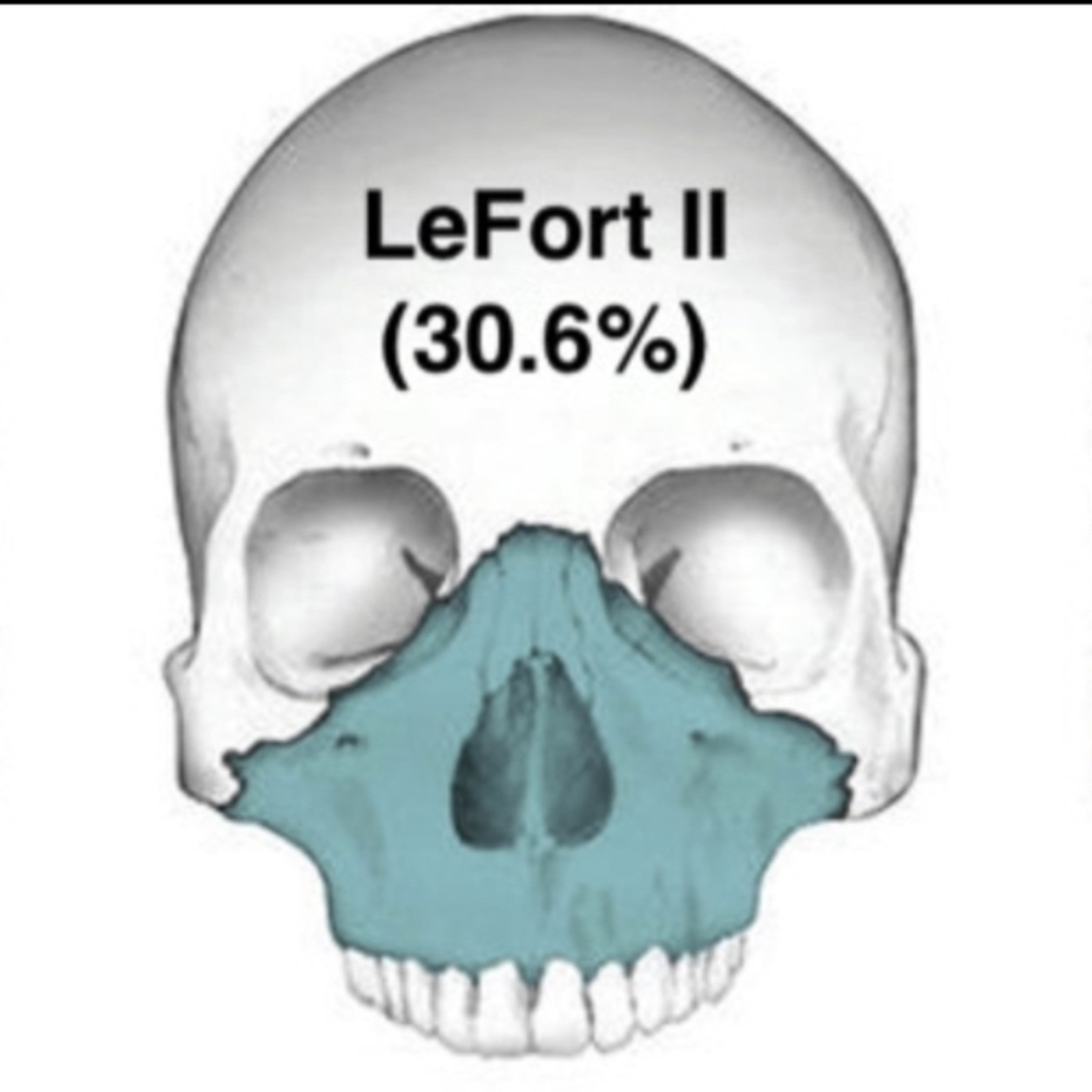
12.23.22 Quick Review #84 - #trauma #facialtrauma #lefort #fracture #fractures #maxillofacial #maxillofacialsurgery #oralandmaxillofacialsurgery #omfs #surgery #surgeon #doctorgallagher #oralsurgery #oralsurgeon #dentist #dentistry #dental LeFort fractures, initially described by René Le Fort in 1901, are a type of facial fracture that involve the maxillary bone and are classified into three main types, based on the level of fracture and the parts of the face they affect: 1. LeFort I Fracture: This is a horizontal fracture that traverses above the alveolar ridge and teeth, from the piriform aperture, along the lateral walls of the maxillary antrum, and extend posteriorly to involve and separate the pterygoid plates. Medially they pass along the lateral nasal wall and the lower third of the septum. These fractures allow the maxillae and hard palate to move as a single block. It is possible to have a unilateral fracture at this level. This would often involve separation along the mid palate. 2. LeFort II Fracture: Also known as a pyramidal fracture, this type involves most of the nasal bones, the maxillary bones, the palatine bones, and the lower two-thirds of the nasal septum, the dentoalveolar structures and the pterygoid plates. The fracture disconnexts superiorly at the nasofrontal junction and continues along the medial inferior third of the orbit and laterally along the zygomaticomaxillary suture and on toward the pterygoid plates. The nasal septum is separated superiorly causing a pyramidal disjunction of the midface. 3. LeFort III Fracture: This is the most severe type. The fractures run at the midline either across the nasal bones or disjoint at the nasofrontal junction, laterally traversing through the medial orbital wall and the superior orbit extending along the inferior orbital fissure and the lateral orbital wall to the zygomaticofrontal suture. The zygomaticotemporal suture is separated as well. The septum separates at the cribriform ethmoid plate. Thus, in the LeFort III fracture type, there is a separation of all of the facial bones from the cranial base. Each type of LeFort fracture can have significant implications for the patient, often requiring surgical intervention to repair and realign the bones. They are commonly the result of high-impact trauma, such as in motor vehicle accidents. References: 1. Patel, B. C., Wright, T., & Waseem, M. (2023). LeFort Fractures. In StatPearls, StatPearls Publishing. 2. “LeFort III fractures: An approach to resuscitation and management” on PubMed. 3. Abubaker, A.O., Lam, D., & Benson, K. (2016). OMFS Secrets (3rd ed.). Elsevier. 4. AO Foundation. (n.d.). AO Surgery Reference. Retrieved from https://lnkd.in/effrrBzH 5. ChatGPT. 2023.
What this episode covers
12.23.22 Quick Review #84 - #trauma #facialtrauma #lefort #fracture #fractures #maxillofacial #maxillofacialsurgery #oralandmaxillofacialsurgery #omfs #surgery #surgeon #doctorgallagher #oralsurgery #oralsurgeon #dentist #dentistry #dental LeFort fractures, initially described by René Le Fort in 1901, are a type of facial fracture that involve the maxillary bone and are classified into three main types, based on the level of fracture and the parts of the face they affect: 1. LeFort I Fracture: This is a horizontal fracture that traverses above the alveolar ridge and teeth, from the piriform aperture, along the lateral walls of the maxillary antrum, and extend posteriorly to involve and separate the pterygoid plates. Medially they pass along the lateral nasal wall and the lower third of the septum. These fractures allow the maxillae and hard palate to move as a single block. It is possible to have a unilateral fracture at this level. This would often involve separation along the mid palate. 2. LeFort II Fracture: Also known as a pyramidal fracture, this type involves most of the nasal bones, the maxillary bones, the palatine bones, and the lower two-thirds of the nasal septum, the dentoalveolar structures and the pterygoid plates. The fracture disconnexts superiorly at the nasofrontal junction and continues along the medial inferior third of the orbit and laterally along the zygomaticomaxillary suture and on toward the pterygoid plates. The nasal septum is separated superiorly causing a pyramidal disjunction of the midface. 3. LeFort III Fracture: This is the most severe type. The fractures run at the midline either across the nasal bones or disjoint at the nasofrontal junction, laterally traversing through the medial orbital wall and the superior orbit extending along the inferior orbital fissure and the lateral orbital wall to the zygomaticofrontal suture. The zygomaticotemporal suture is separated as well. The septum separates at the cribriform ethmoid plate. Thus, in the LeFort III fracture type, there is a separation of all of the facial bones from the cranial base. Each type of LeFort fracture can have significant implications for the patient, often requiring surgical intervention to repair and realign the bones. They are commonly the result of high-impact trauma, such as in motor vehicle accidents. References: 1. Patel, B. C., Wright, T., & Waseem, M. (2023). LeFort Fractures. In StatPearls, StatPearls Publishing. 2. “LeFort III fractures: An approach to resuscitation and management” on PubMed. 3. Abubaker, A.O., Lam, D., & Benson, K. (2016). OMFS Secrets (3rd ed.). Elsevier. 4. AO Foundation. (n.d.). AO Surgery Reference. Retrieved from https://lnkd.in/effrrBzH 5. ChatGPT. 2023.
NOW PLAYING
“What are the 3 LeFort Fracture Types?”
No transcript for this episode yet
Similar Episodes
Dec 5, 2025 ·50m
Oct 9, 2025 ·33m
Oct 3, 2025 ·40m
Sep 11, 2025 ·31m
Aug 27, 2025 ·39m
Aug 18, 2025 ·54m