EPISODE · Jan 16, 2025 · 2 MIN
📝 “What are the Adenomatoid Odontogenic Tumor Variants?”
from Dr. Gallagher's Podcast · host Brendan Gallagher, DDS
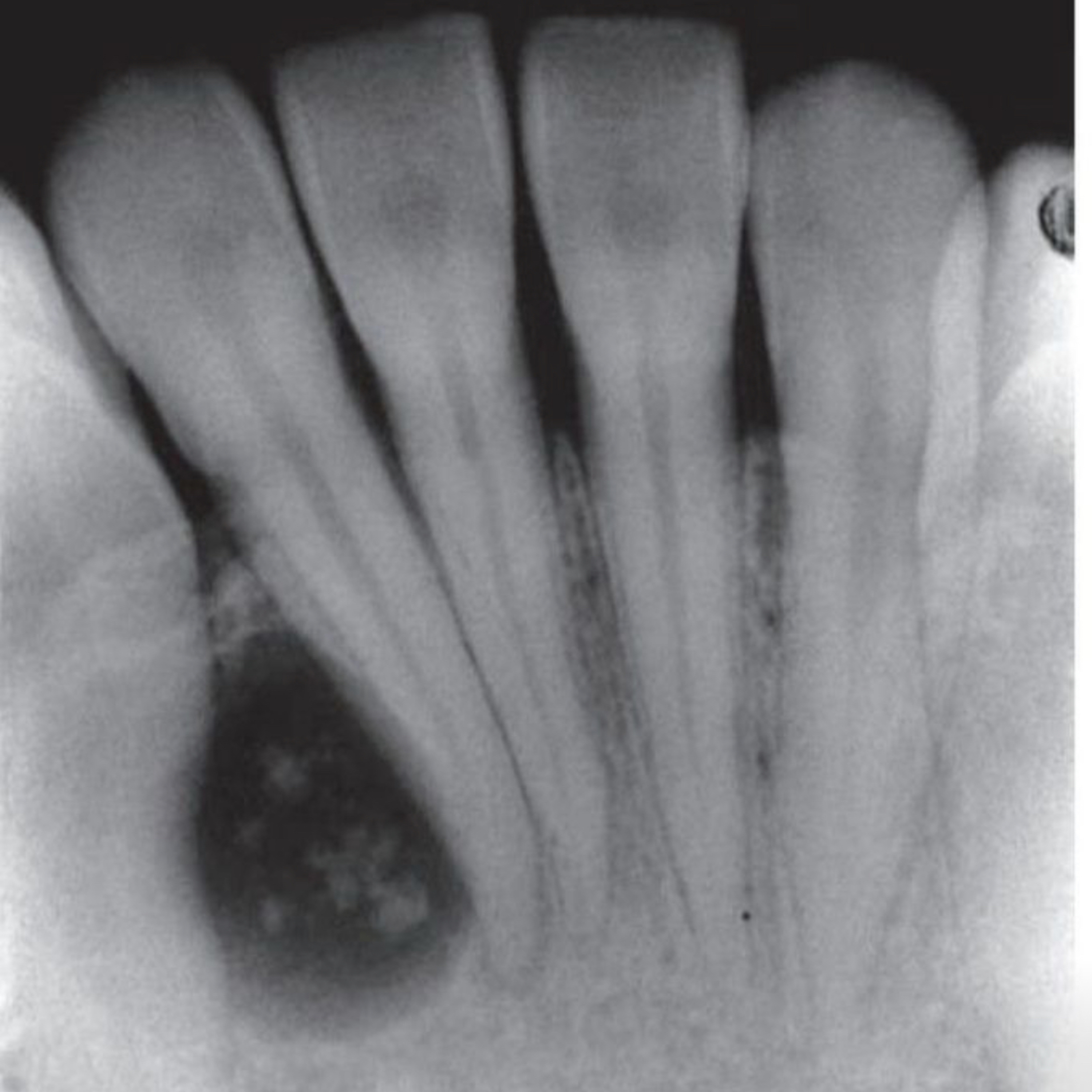
- 1.16.25Quick Review #264 - #pathology #oralpathology #doctorgallagher #oralsurgery #oralsurgeon #dentist #dentistry #dental The adenomatoid odontogenic tumor (AOT) is a benign epithelial odontogenic tumor, slow-growing and well-demarcated. The AOT is classified into three variants: follicular, extrafollicular, and peripheral.1. Follicular Variant• Definition: The most common form, associated with the crown of an unerupted tooth, often a maxillary canine.• Key Features:• Radiographically, it appears as a well-defined radiolucency around the crown, extending along the root.• Mimics a dentigerous cyst but shows tumor-like behavior.• Histology reveals duct-like epithelial structures, spindle-shaped cells, and calcifications.• Differentiation: Distinguished by its tumor-like features and the presence of epithelial duct-like formations.2. Extrafollicular Variant• Definition: Occurs independently of unerupted teeth, commonly in the anterior maxilla.• Key Features:• Appears as a solitary radiolucency with varying degrees of radiopacity from calcifications.• Not associated with specific teeth but can cause cortical expansion.• Histologically identical to the follicular variant.• Differentiation: Defined by the absence of association with an unerupted tooth.3. Peripheral Variant• Definition: The least common, arising in the gingival soft tissues.• Key Features:• Appears as a small gingival mass with minimal or no radiographic changes.• Histologically consistent with other variants, featuring duct-like structures and calcifications.References:1. Saleh, Z. (n.d.). Periapical radiograph of anterior mandibular incisors showing radiolucency. Retrieved from https://lnkd.in/dPR3SstP2. Neville, B. W., Damm, D. D., Allen, C. M., & Chi, A. C. (2015). Oral and Maxillofacial Pathology (4th ed.). Saunders.3. Philipsen, H. P., & Reichart, P. A. (1998). Adenomatoid odontogenic tumor: Facts and figures. Oral Oncology, 35(2), 125-131. https://lnkd.in/dztuvQF34. Eversole, L. R. (2008). Clinical outline of oral pathology: Diagnosis and treatment (4th ed.). BC Decker.5. ChatGPT. 2025.#podcast #dentalpodcast #doctorgallagherpodcast #doctorgallagherspodcast #doctor #dentist #dentistry #oralsurgery #dental #dentalschool #dentalstudent #doctorlife #dentistlife #oralsurgeon #doctorgallagher
What this episode covers
- 1.16.25Quick Review #264 - #pathology #oralpathology #doctorgallagher #oralsurgery #oralsurgeon #dentist #dentistry #dental The adenomatoid odontogenic tumor (AOT) is a benign epithelial odontogenic tumor, slow-growing and well-demarcated. The AOT is classified into three variants: follicular, extrafollicular, and peripheral.1. Follicular Variant• Definition: The most common form, associated with the crown of an unerupted tooth, often a maxillary canine.• Key Features:• Radiographically, it appears as a well-defined radiolucency around the crown, extending along the root.• Mimics a dentigerous cyst but shows tumor-like behavior.• Histology reveals duct-like epithelial structures, spindle-shaped cells, and calcifications.• Differentiation: Distinguished by its tumor-like features and the presence of epithelial duct-like formations.2. Extrafollicular Variant• Definition: Occurs independently of unerupted teeth, commonly in the anterior maxilla.• Key Features:• Appears as a solitary radiolucency with varying degrees of radiopacity from calcifications.• Not associated with specific teeth but can cause cortical expansion.• Histologically identical to the follicular variant.• Differentiation: Defined by the absence of association with an unerupted tooth.3. Peripheral Variant• Definition: The least common, arising in the gingival soft tissues.• Key Features:• Appears as a small gingival mass with minimal or no radiographic changes.• Histologically consistent with other variants, featuring duct-like structures and calcifications.References:1. Saleh, Z. (n.d.). Periapical radiograph of anterior mandibular incisors showing radiolucency. Retrieved from https://lnkd.in/dPR3SstP2. Neville, B. W., Damm, D. D., Allen, C. M., & Chi, A. C. (2015). Oral and Maxillofacial Pathology (4th ed.). Saunders.3. Philipsen, H. P., & Reichart, P. A. (1998). Adenomatoid odontogenic tumor: Facts and figures. Oral Oncology, 35(2), 125-131. https://lnkd.in/dztuvQF34. Eversole, L. R. (2008). Clinical outline of oral pathology: Diagnosis and treatment (4th ed.). BC Decker.5. ChatGPT. 2025.#podcast #dentalpodcast #doctorgallagherpodcast #doctorgallagherspodcast #doctor #dentist #dentistry #oralsurgery #dental #dentalschool #dentalstudent #doctorlife #dentistlife #oralsurgeon #doctorgallagher
NOW PLAYING
📝 “What are the Adenomatoid Odontogenic Tumor Variants?”
No transcript for this episode yet
Similar Episodes
Dec 5, 2025 ·50m
Oct 9, 2025 ·33m
Oct 3, 2025 ·40m
Sep 11, 2025 ·31m
Aug 27, 2025 ·39m
Aug 18, 2025 ·54m