PODCAST · science
Light Sheet Sheds Light on Tumor Therapy
by Svenja Rühland / Hartmann Harz
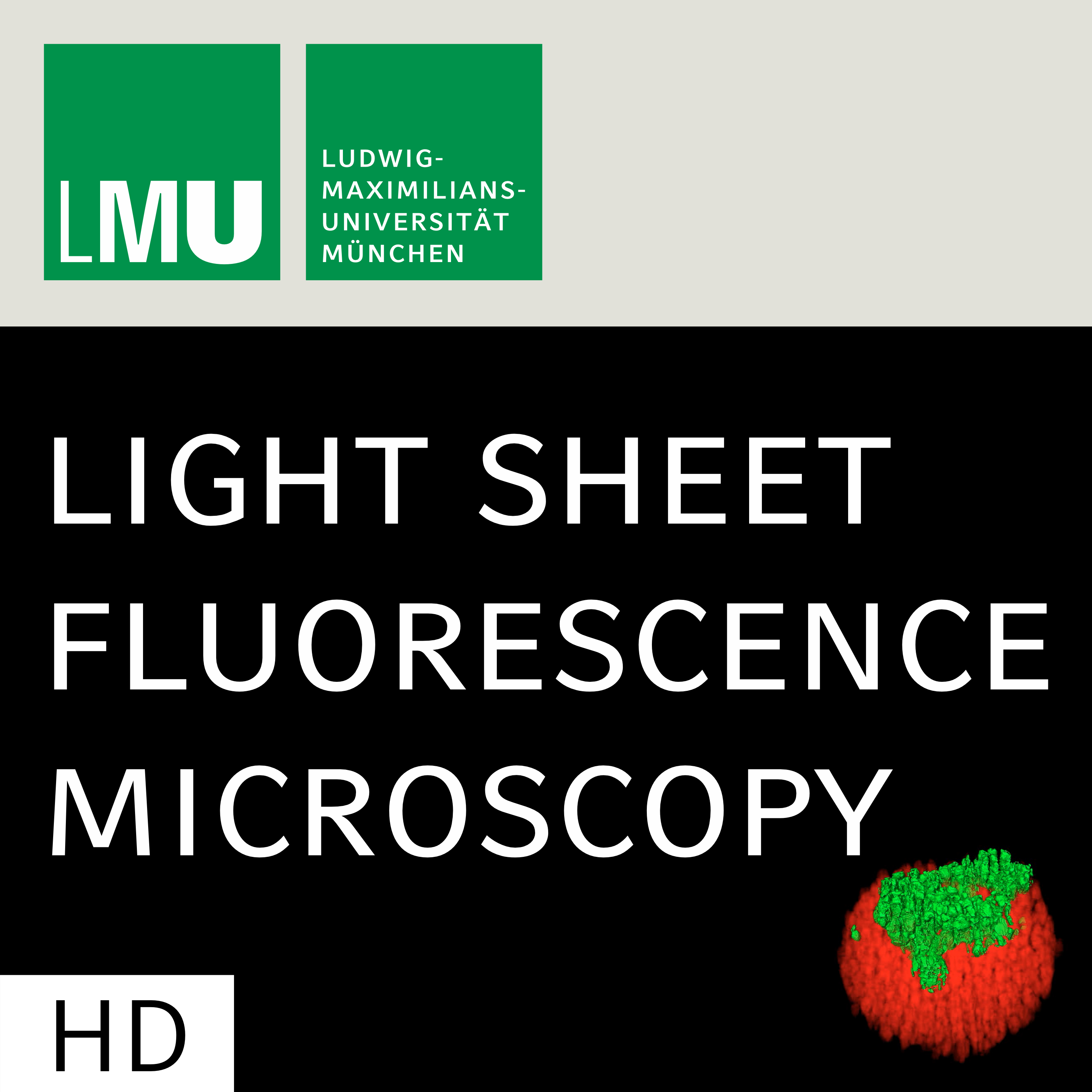
The idea of light sheet microscopy was already born a hundred years ago. Richard Zsigmondy used an innovative method of side illumination to observe the chemistry of nanometer sized colloids, which won him the Nobel Prize in chemistry in 1925. Almost a century later, Ernst Stelzer and his group re-implemented that technology within an era dominated by fluorescence microscopy. Fluorescent labeling of cells and the sensitivity of light sheet microscopy led to new insights into the three-dimensionality of whole organs and the four-dimensionality of developing organisms. The open source project “openSPIM” by the group of Pavel Tomancak finally made light sheet microscopy accessible to the broad scientific community. This has allowed Hartmann Harz at the Center for Advanced Light Microscopy at the LMU Munich to build an in-house light sheet microscope which PhD student Svenja Rühland from the group of Peter Nelson uses to image therapeutic vehicle cells within 3D tumor spheroids.
-
1
Light Sheet Sheds Light on Tumor Therapy (CC)
The idea of light sheet microscopy was already born a hundred years ago. Richard Zsigmondy used an innovative method of side illumination to observe the chemistry of nanometer sized colloids, which won him the Nobel Prize in chemistry in 1925. Almost a century later, Ernst Stelzer and his group re-implemented that technology within an era dominated by fluorescence microscopy. Fluorescent labeling of cells and the sensitivity of light sheet microscopy led to new insights into the three-dimensionality of whole organs and the four-dimensionality of developing organisms. The open source project “openSPIM” by the group of Pavel Tomancak finally made light sheet microscopy accessible to the broad scientific community. This has allowed Hartmann Harz at the Center for Advanced Light Microscopy at the LMU Munich to build an in-house light sheet microscope which PhD student Svenja Rühland from the group of Peter Nelson uses to image therapeutic vehicle cells within 3D tumor spheroids.
We're indexing this podcast's transcripts for the first time — this can take a minute or two. We'll show results as soon as they're ready.
No matches for "" in this podcast's transcripts.
No topics indexed yet for this podcast.
Loading reviews...
ABOUT THIS SHOW
The idea of light sheet microscopy was already born a hundred years ago. Richard Zsigmondy used an innovative method of side illumination to observe the chemistry of nanometer sized colloids, which won him the Nobel Prize in chemistry in 1925. Almost a century later, Ernst Stelzer and his group re-implemented that technology within an era dominated by fluorescence microscopy. Fluorescent labeling of cells and the sensitivity of light sheet microscopy led to new insights into the three-dimensionality of whole organs and the four-dimensionality of developing organisms. The open source project “openSPIM” by the group of Pavel Tomancak finally made light sheet microscopy accessible to the broad scientific community. This has allowed Hartmann Harz at the Center for Advanced Light Microscopy at the LMU Munich to build an in-house light sheet microscope which PhD student Svenja Rühland from the group of Peter Nelson uses to image therapeutic vehicle cells within 3D tumor spheroids.
HOSTED BY
Svenja Rühland / Hartmann Harz
Loading similar podcasts...
Frequently Asked Questions
How many episodes does Light Sheet Sheds Light on Tumor Therapy have?
Light Sheet Sheds Light on Tumor Therapy currently has 1 episodes available on PodParley. New episodes are automatically indexed when they're published to the podcast feed.
What is Light Sheet Sheds Light on Tumor Therapy about?
The idea of light sheet microscopy was already born a hundred years ago. Richard Zsigmondy used an innovative method of side illumination to observe the chemistry of nanometer sized colloids, which won him the Nobel Prize in chemistry in 1925. Almost a century later, Ernst Stelzer and his group...
How often does Light Sheet Sheds Light on Tumor Therapy release new episodes?
Light Sheet Sheds Light on Tumor Therapy has 1 episodes. Check the episode list to see recent publication dates and frequency.
Where can I listen to Light Sheet Sheds Light on Tumor Therapy?
You can listen to Light Sheet Sheds Light on Tumor Therapy on PodParley by clicking any episode. We provide an embedded audio player for direct listening, and you can also subscribe via your preferred podcast app using the RSS feed.
Who hosts Light Sheet Sheds Light on Tumor Therapy?
Light Sheet Sheds Light on Tumor Therapy is created and hosted by Svenja Rühland / Hartmann Harz.
URL copied to clipboard!