
EPISODE · Jul 17, 2025 · 8 MIN
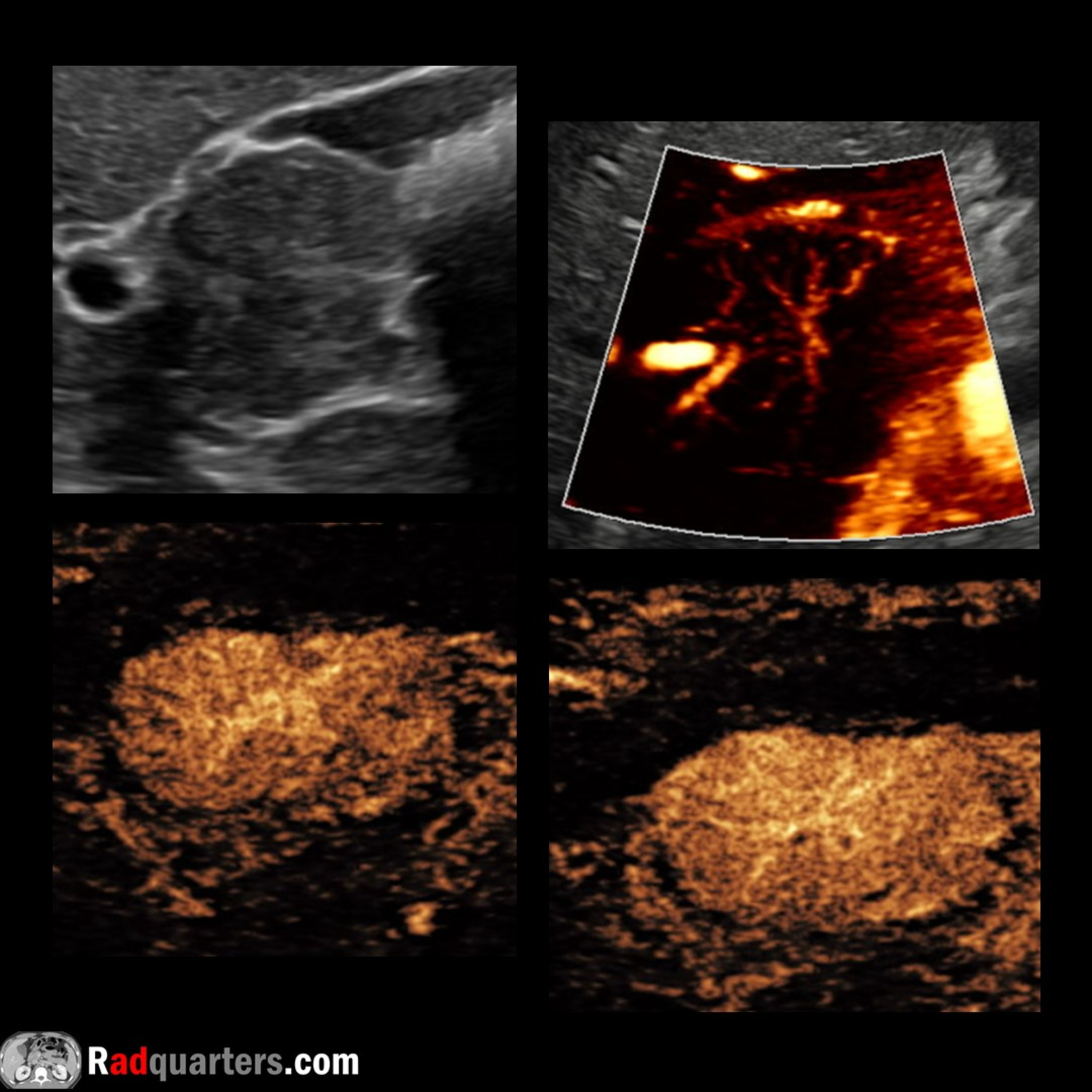
Contrast-Enhanced Ultrasound of Focal Nodular Hyperplasia
from Radiology Lectures | Radquarters · host Daniel J. Kowal, MD
In this radiology lecture, we review the ultrasound and contrast-enhanced ultrasound appearance of focal nodular hyperplasia!Key teaching points include:Focal nodular hyperplasia (FNH) is the second most common benign liver tumor after hemangiomaMost common in women of reproductive age with highest incidence between the ages of 20-50. Female to male predilection 8:1No association with oral contraceptives (unlike hepatic adenoma)No risk of malignant potential, and no surgery required = A leave alone lesionUnclear etiology, thought to be a regenerative, hyperplastic response of normal hepatocytes to anomalous arteries within liverHamartomatous-type lesion containing dense collection of functional hepatocytes and malformed, blind-ending biliary ductules leading to slowed biliary excretionFunctioning Kupffer cells (hepatic macrophages) may be presentUS appearance variable: Can be hypoechoic, isoechoic or hyperechoic relative to liver parenchyma. May have an echogenic central scarSpoke-wheel sign of central vascularity is a helpful ancillary feature, and seen in up to 63% of FNH with microflow imaging*FNH appearance on contrast-enhanced ultrasound (CEUS): Immediate stellate enhancement radiating from the center of the lesion in centrifugal fashion, followed by diffuse homogeneous hyperenhancement relative to the surrounding parenchymaCEUS has high specificity in the diagnosis of FNH**References*Kang TW, et al. Comparison of Super-Resolution US and Contrast Material-enhanced US in Detection of the Spoke Wheel Sign in Patients with FNH. Radiology. 2021;298(1):82-90**Pei, X-Q et al. Quantitative analysis of contrast-enhanced ultrasonography: differentiating focal nodular hyperplasia from hepatocellular carcinoma. Br J Radiol. 2013 Mar;86(1023):20120536To learn more about the Samsung RS85 Prestige ultrasound system, please visit: https://www.bostonimaging.com/rs85-prestige-ultrasound-system-4Click the YouTube posts tab or follow on social media for bonus teaching material posted throughout the week!Website: https://radquarters.com/Instagram: https://www.instagram.com/radquarters/Facebook: https://www.facebook.com/radquarters/X (Twitter): https://twitter.com/radquartersReddit: https://www.reddit.com/user/radiologistHQ/This video is for informational purposes only. It does not replace the advice or counsel of a doctor or health care professional.
Embed this episode
NOW PLAYING
Contrast-Enhanced Ultrasound of Focal Nodular Hyperplasia
No transcript for this episode yet
Similar Episodes
No similar episodes found.
Similar Podcasts
No similar podcasts found.