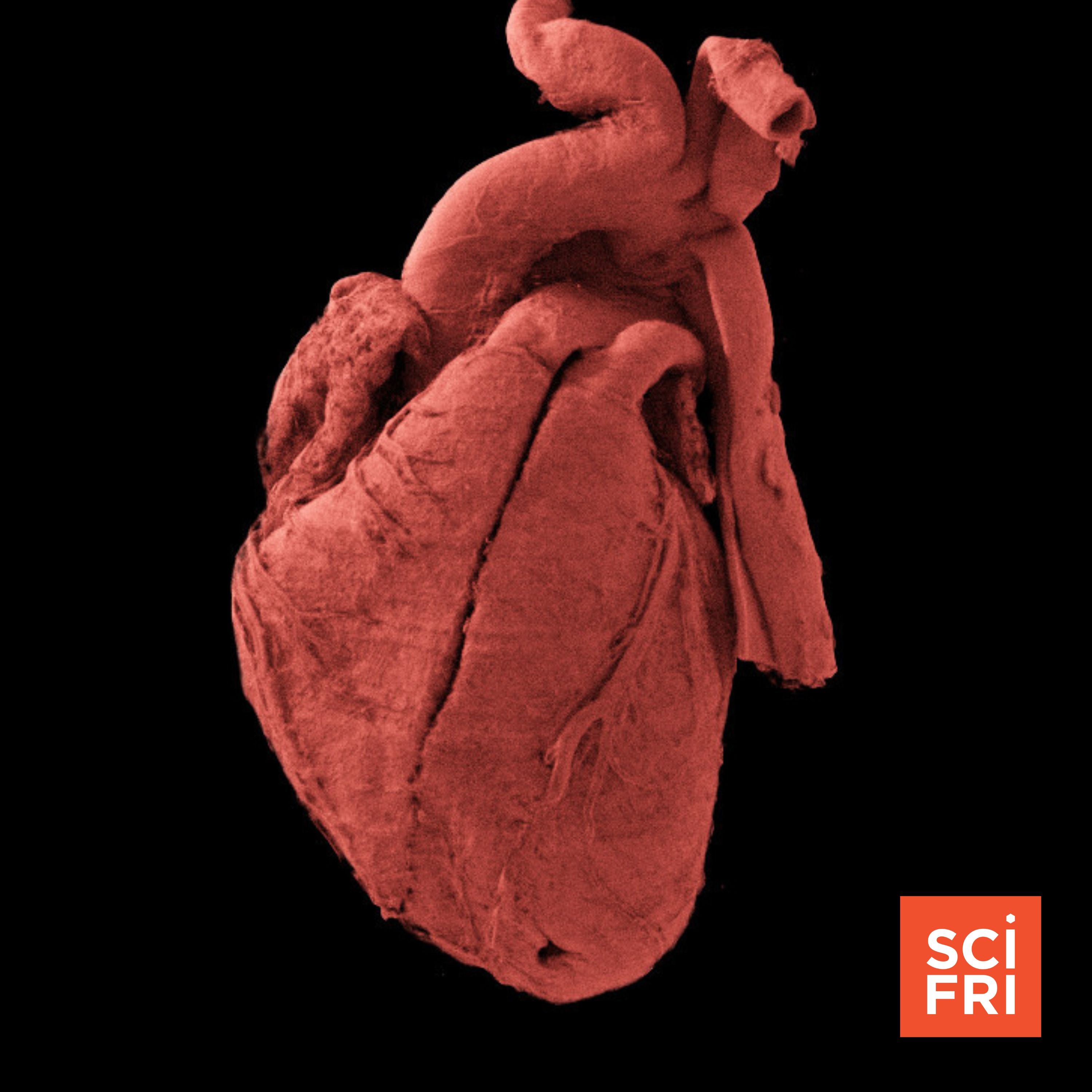
How a particle accelerator illuminated 56 human organs
from Science Friday Hosted by Dee Peterschmidt, Ira Flatow, Claire Walsh
A new imaging technique using a particle accelerator is giving researchers an unprecedented level of detail of our organs, producing scans 100 billion times brighter than a CT scanner. Those 3D models are now part of a public database called the Human Organ Atlas, available to researchers and the medically curious. Joining Host Ira Flatow to explain why they needed so much power and what kind of research advances will follow is imaging scientist Claire Walsh, director of the Human Organ Atlas hub. Check out images from the Human Organ Atlas on our website. Guest: Dr. Claire Walsh is an associate professor at the UCL department of mechanical engineering and director of the Human Organ Atlas Hub. Transcripts for each episode are available within 1-3 days at sciencefriday.com. Subscribe to this podcast. Follow us on socials @ scifri on YouTube, Instagram, Facebook, TikTok, and Bluesky and sign up for our newsletters. Got a science question that’s keeping you up at night? Call us: 877-472-4374
Embed this episode
What this episode covers
The Human Organ Atlas gives an extremely detailed look at 56 human organs, scanned with the help of a particle accelerator.
No transcript yet.