
EPISODE · Aug 18, 2025 · 4 MIN
"How Do You Manage Bleeding During A Le Fort I Osteotomy?"
from Dr. Gallagher's Podcast · host Brendan Gallagher, DDS
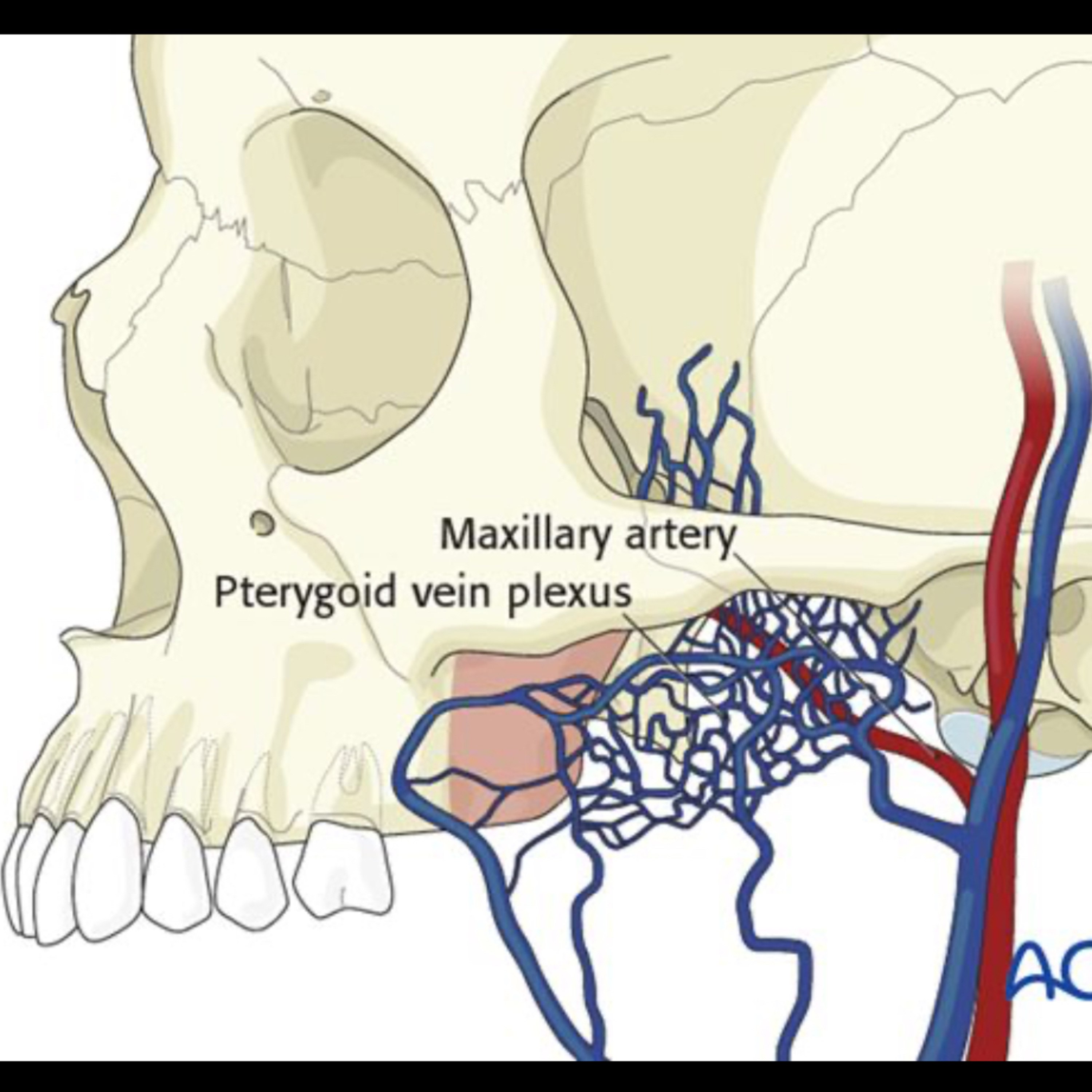
Quick Review #280 - #surgery #surgeon #doctorgallagher #oralsurgery #oralsurgeon #omfs #dentist #dentistry #dental #orthognathic #lefort #bsso #bleeding - 8.17.25 Bleeding during a Le Fort I osteotomy, particularly at the pterygomaxillary junction, is a high-yield oral boards topic and a common intraoperative challenge. Frequent sources are the descending palatine artery (arterial) and the pterygoid venous plexus (venous). Anatomy & Mechanism: The descending palatine artery, a branch of the maxillary artery, travels through the greater palatine canal, emerging at the posterior hard palate. It lies close to the posterior maxillary wall and is vulnerable when separating the maxilla from the pterygoid plates. The pterygoid venous plexus is located around the lateral pterygoid muscle and produces slow, dark venous oozing rather than pulsatile bleeding. Recognition: A) Arterial bleeding: Bright red, pulsatile flow; rapid accumulation in the field. B) Venous bleeding: Dark, slow ooze; diffuse origin. Immediate Management: 1. Direct Pressure – Apply firm pressure with gauze or sponge at the bleeding point. 2. Suction & Visualization – Clear the field to localize the source. 3. Hemostatic Agents – Surgicel, Gelfoam, or Floseal for diffuse venous ooze. 4. Electrocautery – Bipolar cautery for focal arterial injury, avoiding nerve injury (e.g., V2 branches). 5. Packing – If uncontrollable, pack the area temporarily and assess for stability. 6. Escalation – For severe or persistent arterial bleeding, consider intraoperative ligation or postoperative embolization of the maxillary artery via interventional radiology. Prevention: 1. Keep osteotomes oriented correctly during posterior maxillary cuts. 2. Avoid excessive posterior force that risks the greater palatine canal. 3. Use virtual surgical planning to anticipate anatomy and avoid injury zones. Tip: Try to identify whether the bleed is arterial or venous—management and urgency might differ. Mention both possible sources in your oral boards answer, but highlight the descending palatine artery as the more common significant arterial injury in Le Fort I procedures. References: 1. Bartlett, S., Ehrenfeld, M., Mast, G., & Sugar, A. (n.d.). Approach to the Le Fort I level of the midface in cleft lip and palate patients. In E. Ellis III (Exec. Ed.) & D. Buchbinder (Gen. Ed.), AO Surgery Reference: CMF – Congenital Deformities. AO Foundation. Retrieved August 14, 2025, from https://lnkd.in/dvQj9tr3 2. Miloro, M., Ghali, G. E., Larsen, P. E., & Waite, P. (2022). Peterson’s Principles of Oral and Maxillofacial Surgery (4th ed.). Springer. 3. Abubaker, A. O., Lam, D., & Benson, K. (2016). Oral and Maxillofacial Surgery Secrets (3rd ed.). Elsevier. 4. ChatGPT. 2025 #podcast #dentalpodcast #doctorgallagherpodcast #doctorgallagherspodcast #doctor #dentist #dentistry #oralsurgery #dental #dentalschool #dentalstudent #doctorlife #dentistlife #oralsurgeon #doctorgallagher
Embed this episode
Ready to play
"How Do You Manage Bleeding During A Le Fort I Osteotomy?"
No transcript for this episode yet
Similar Episodes
No similar episodes found.
Similar Podcasts
No similar podcasts found.