
EPISODE · Sep 13, 2026 · 45 MIN
PSYCH 121: Anxiety Disorders - Neuroimaging and Neuroanatomical Circuits
from Clinical Deep Dives · host Dr Manaan Kar Ray
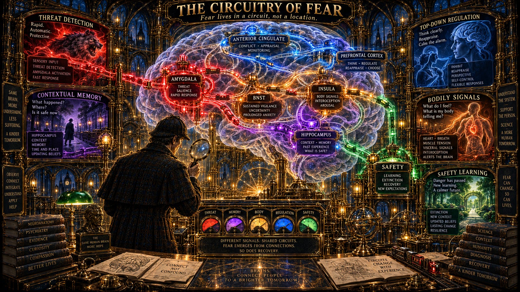
Medlock Holmes enters the Grand Neuroanatomical Theatre of Fear.At the centre hangs an enormous transparent brain.The amygdala glows brightly.But Holmes does not stop there.Around it are the hippocampus.The medial and orbital prefrontal cortex.The anterior cingulate cortex.The insula.The thalamus.The hypothalamus.The bed nucleus of the stria terminalis.The periaqueductal grey.The locus coeruleus.Sensory association cortices.White-matter pathways.Every structure is connected.The chapter begins with an important methodological principle.To understand the circuitry of anxiety, investigators move step by step:First, identify the clinical phenomenon.Then identify the brain systems that normally perform that function.Then determine whether those systems behave differently in people with anxiety disorders.Finally, ask whether those abnormalities relate to symptoms, course, or treatment response.The functional problems are familiar:Persistent learning about threat.Exaggerated fear.Poor extinction.Difficulty suppressing attention to danger.Overgeneralisation from one threat to similar safe stimuli.Heightened perception of threat.Increased sensitivity to bodily sensations.Neuroimaging attempts to map these phenomena onto circuits.Holmes enters the Fear Learning Laboratory.A neutral cue appears.Then an aversive event.Again.And again.Eventually the cue alone produces fear.The cue has become the CS+.Another cue that was never paired with danger becomes the CS−.The amygdala learns the association.But then the experiment changes.The CS+ is repeatedly presented without the aversive event.Fear gradually decreases.This is extinction.Holmes notices something crucial.Extinction does not erase the original fear memory.It creates a new competing safety memory.That means extinguished fear can return.After time.In another context.After an unexpected aversive event.The fear memory was not destroyed.It was inhibited.The chapter distinguishes this from memory reconsolidation.When a fear memory is retrieved, it temporarily becomes unstable.During a reconsolidation window, new information may modify the original memory itself.This raises the possibility that future interventions might not simply suppress old fear, but update it.The clinical relevance is immediate.Exposure therapy works through these same learning principles.The therapeutic challenge is therefore not merely helping the patient endure fear.It is helping the brain encode and retain safety.Holmes now enters the Amygdala Chamber.Sensory information arrives through two major routes.The first is fast.The thalamus sends relatively crude information directly towards the lateral amygdala.Danger can be detected before the cortex has fully analysed what is happening.The second is slower.Sensory cortex processes the stimulus in greater detail before communicating with the amygdala.The lateral amygdala receives converging information about the conditioned and unconditioned stimuli.The basal and accessory basal nuclei help shape learning and memory.The central nucleus becomes the major output station.From there, signals descend towards the hypothalamus, brainstem, and motor systems.Heart rate rises.Blood pressure changes.Breathing accelerates.The body freezes or prepares to flee.Stress hormones are released.Facial expression changes.The figure on page 6 maps this architecture beautifully: external threat enters through sensory and thalamic pathways, converges upon the amygdala, and then recruits systems controlling endocrine, autonomic, behavioural, and motor responses.But Holmes notices another structure beside the amygdala:The Bed Nucleus of the Stria Terminalis.The distinction becomes fundamental.The amygdala is strongly involved in acute fear.The BNST is particularly important for sustained anxiety.An obvious danger appears.The amygdala responds.An uncertain threat may appear sometime soon.The BNST keeps the organism vigilant.This distinction becomes especially important in generalized anxiety disorder.Holmes then reaches the Prefrontal Control Room.The medial prefrontal cortex and anterior cingulate are not passive observers.They regulate fear.Interpret context.Predict consequences.Modify behaviour.Help determine whether the amygdala should continue sounding the alarm.The prelimbic and infralimbic regions in animal models provide an important conceptual framework.Prelimbic circuitry can support fear expression.Infralimbic circuitry contributes to extinction.In humans, the ventromedial prefrontal cortex performs an analogous regulatory role.The source’s figure on page 9 places fear expression and fear extinction beside one another.During fear expression, amygdala output activates downstream physiological responses.During extinction, hippocampal contextual information and infralimbic/vmPFC pathways recruit inhibitory interneurons within the amygdala, suppressing central-amygdala output.The same fear cue can therefore produce a very different response depending on which circuit dominates.Holmes sees anxiety as a problem of balance.Threat detection must be fast enough to protect.But cortical regulation must be strong enough to stop fear when the danger is no longer relevant.The next chamber belongs to the hippocampus.The amygdala asks:“Is this dangerous?”The hippocampus adds:“Where are we?”Context matters.A person bitten by a dog may learn fear.But whether that fear is expressed later depends partly on whether the current environment resembles the original context.The hippocampus helps distinguish:This place was dangerous.This place is safe.This cue used to predict harm.Here, it no longer does.Without contextual discrimination, fear generalises.The same signal spreads across places and situations that were never dangerous.The hippocampus therefore helps determine whether the original fear memory or the newer extinction memory should dominate.Holmes then encounters the Orbitofrontal Chamber.The orbitofrontal cortex tracks changing reinforcement.What used to predict danger may no longer do so.What used to be safe may now carry risk.Behaviour must change accordingly.Damage to orbitofrontal systems produces perseveration.The person continues using an old strategy despite changed circumstances.In anxiety, dysfunctional orbitofrontal regulation may contribute to continued fearful thoughts and behaviours even when reinforcement no longer supports them.The next room belongs to the insula.Here, Holmes hears the body.Heartbeat.Breathing.Nausea.Chest tightness.Internal temperature.Visceral sensations.The insula plays a central role in interoception.This is particularly relevant to panic disorder.The person does not merely experience bodily sensations.They monitor them.Interpret them.Amplify them.A harmless change in heart rate becomes evidence of catastrophe.The brain’s representation of internal bodily state becomes part of the threat itself.Holmes enters the Imaging Observatory.Different instruments examine different dimensions of brain structure and function.Structural MRI measures regional size and shape.Voxel-based morphometry compares grey-matter architecture.Diffusion tensor imaging estimates white-matter organisation through measures such as fractional anisotropy.PET examines blood flow or metabolism.fMRI measures BOLD signal.EEG records electrical activity.MEG measures magnetic fields generated by neuronal activity.But Holmes quickly learns that brain imaging is always state dependent.A resting brain.A brain viewing fearful faces.A brain recalling panic.A brain anticipating shock.A brain undergoing treatment.These are not equivalent experiments.Neuroimaging therefore depends as much on the task as on the scanner.The next chamber examines healthy fear learning.Human studies repeatedly show increased amygdala activity to a CS+ compared with a CS−.During extinction, vmPFC activation becomes important.Stronger vmPFC activation predicts better retention of extinction.The hippocampus becomes particularly active when context determines whether fear or safety should be expressed.Together, the amygdala, vmPFC, and hippocampus form a central circuit for human fear acquisition and extinction.Holmes then enters the Emotional Face Gallery.Hundreds of faces look towards him.Fearful.Angry.Happy.Neutral.Surprised.The amygdala responds to many emotionally salient expressions, but fearful and ambiguous faces can produce particularly strong responses.Why?A fearful face says:“Something dangerous is nearby.”But it does not reveal what the danger is.Uncertainty itself becomes salient.Even when fearful faces are presented too briefly for conscious recognition, the amygdala can still respond.Threat processing can therefore occur before explicit awareness.The anterior cingulate detects conflict.The lateral prefrontal cortex helps redirect attention.The insula monitors bodily state.The amygdala enhances memory for emotionally salient events.Holmes sees why anxiety can become so persistent.The brain not only detects threat.It can preferentially remember it.The investigation now turns to individual disorders.The first chamber is Panic Disorder.Holmes must explain something unusual.Why does panic sometimes appear to occur without any trigger?One theory suggests a false alarm.Normal fear circuitry activates inappropriately because of abnormalities in internal homeostatic systems.Another theory suggests failed regulation.A small anxiety response begins but cortical systems fail to contain it.A third possibility is that the trigger exists, but is processed outside conscious awareness.The patient experiences the attack as spontaneous.The brain may have detected something the conscious mind did not.Neuroimaging in panic disorder shows abnormalities across hippocampal and parahippocampal regions, temporal cortex, amygdala, insula, ACC, orbitofrontal cortex, and prefrontal systems.Resting-state abnormalities are found even when no panic attack is occurring.During panic-related provocation, the insula, frontal cortex, cingulate cortex, hippocampus, and striatum may become unusually active.Emotional-face studies often show increased amygdala and insula activation alongside reduced vmPFC and ACC recruitment.The overall pattern suggests excessive bottom-up alarm combined with insufficient top-down regulation.Treatment begins to alter this architecture.Following CBT, activity in frontal regions can decrease while functional connectivity with the amygdala, hippocampus, and prefrontal systems improves.Stronger baseline negative ACC–amygdala connectivity has even been associated with better later response to pharmacotherapy or CBT.Holmes writes:Treatment does not merely reduce symptoms. It can reorganise the circuitry supporting those symptoms.The next chamber contains Specific Phobias.Here the neurobiology becomes more stimulus-specific.Show a spider to someone with spider phobia.The amygdala activates.The insula activates.The anterior cingulate activates.Present an unrelated emotional stimulus.The same exaggerated response may disappear.The hypersensitivity is closely tied to the feared category.The source describes an elegant distinction.During brief, predictable exposure to a phobic stimulus, the amygdala is particularly active.During prolonged, uncertain threat, the BNST and ACC become more involved.The finding mirrors the larger distinction between immediate fear and sustained anxiety.The source also shows that successful exposure therapy changes brain function.Before treatment:High amygdala.High insula.High ACC activity.Shortly after treatment:Amygdala activation falls.Prefrontal engagement rises.Months later:Amygdala reactivity remains lower, even though the prefrontal system no longer needs to work as hard.The brain has not simply learned to suppress fear consciously.The threat representation itself has become less reactive.Holmes moves next to Social Anxiety Disorder.The feared stimulus is no longer a spider.It is another human being.A face.A judgement.A stare.A critical comment.An audience.Social-anxiety imaging repeatedly shows heightened amygdala responses to socially relevant stimuli.The threshold for perceiving social threat appears lower.The insula also becomes hyperresponsive.Frontolimbic connectivity may be weaker.Most strikingly, the abnormality is selective.A socially anxious patient may show heightened amygdala activity to a human face but not to an aversive smell.Self-referential criticism produces particularly strong amygdala and medial prefrontal responses.The brain is not overreacting equally to everything unpleasant.It is selectively tuned to social evaluation.CBT again changes the circuitry.After treatment, patients show greater dorsolateral and dorsomedial prefrontal engagement during cognitive reappraisal and stronger negative coupling between dmPFC and amygdala.The cortex becomes more effective at regulating limbic response.Pretreatment imaging can even improve prediction of who responds to CBT beyond clinical measures alone.The possibility of personalised psychiatry begins to emerge.The next chamber belongs to Generalized Anxiety Disorder.Here the threat has no single object.The problem is sustained uncertainty.Worry.Overgeneralisation.Failure to distinguish danger from safety.The amygdala findings are therefore less straightforward.Some studies show hyperactivity.Some do not.Some show responses even to neutral stimuli.But the BNST becomes especially important.The source’s figure on page 30 shows delayed but sustained BNST activation during threat anticipation in GAD.The amygdala responds more rapidly.The BNST response emerges later and persists.The brain has moved from:“Danger now.”to“Danger may be coming.”The distinction maps closely onto the clinical difference between fear and anxiety.GAD also shows persistent activity in the dACC and dmPFC after worry induction.Healthy individuals activate these regions during worry, then return towards baseline.People with GAD continue activating them even after the provoking task ends.The worry circuit does not disengage.Fear-generalisation experiments reveal another clue.When a harmless stimulus resembles the original threat cue, people with GAD are more likely to generalise fear towards it.At the same time, vmPFC recruitment is reduced.The brain becomes less effective at saying:“This looks similar, but it is not the danger.”Connectivity between ACC and amygdala is also impaired.The uncinate fasciculus - a major white-matter pathway connecting frontal and temporal regions - shows reduced integrity in some studies.The disorder therefore appears to involve not simply an overactive fear centre, but impaired communication between regulation and threat systems.Holmes now steps back and compares the disorders.The amygdala appears across all of them.But not in the same way.In panic disorder, hyperreactivity may extend across both specific and general threat cues.In specific phobia, the response is particularly tied to the phobic stimulus.In social anxiety, it is especially strong for socially relevant threat.In GAD, the pattern is less consistently amygdala-dominated, with the BNST becoming especially important for sustained anticipatory anxiety.The anterior insula appears across several disorders.The hippocampus seems particularly important in panic disorder.The prefrontal cortex and ACC contribute varying degrees of deficient control.There is therefore both shared circuitry and disorder-specific circuitry.The final major chamber is labelled:DEVELOPMENTHolmes sees the same brain changing across childhood, adolescence, and adulthood.The amygdala develops relatively early.The hippocampus also matures earlier.But prefrontal regulatory systems develop slowly.They do not achieve mature organisation until early adulthood.That creates an important developmental imbalance.The alarm system becomes powerful before the regulatory system has fully matured.Adolescence may therefore represent a particularly vulnerable period for fear regulation.The capacity to extinguish conditioned fear appears reduced during adolescence compared with childhood and adulthood.At the same time, social stimuli become increasingly important.This may help explain why many anxiety disorders peak during adolescence.The source’s figure on page 35 demonstrates something even more important.Amygdala–vmPFC connectivity differs not only by anxiety diagnosis, but by age.Adults and young people with anxiety do not necessarily show the same neural pattern.The biology of anxiety changes across development.That means we cannot simply take a circuit identified in anxious adults and assume it operates identically in children.The developmental context changes everything.Social anxiety provides a striking example.During adolescence, peer evaluation becomes biologically salient.Adolescents with social anxiety show increased amygdala activation when anticipating evaluation from peers they do not want to interact with.They may show reduced nucleus accumbens activation when anticipating feedback from desirable peers.Threat becomes stronger.Reward becomes weaker.The social world becomes organised around potential rejection.In paediatric GAD, amygdala and prefrontal abnormalities are also evident.Some studies suggest increased amygdala reactivity alongside altered vlPFC–amygdala connectivity.The ventrolateral prefrontal cortex may serve a compensatory role.Higher activation can be associated with lower symptom severity.Even treatment response may depend on these developmental circuits.Holmes reaches the final gallery.A child with behavioural inhibition stands before an unfamiliar face.The amygdala responds strongly.Years later, the same pattern may be found in an adult with an anxiety disorder.But Holmes refuses to assume that the abnormality is caused by the illness.Perhaps it existed before the illness.Perhaps it was a risk marker.Perhaps another brain compensates successfully and the disorder never develops.This is one of neuroimaging’s greatest challenges.A difference between patients and controls may represent:Cause.Consequence.Risk.Compensation.Treatment effect.Development.Or an unrelated epiphenomenon.Only longitudinal research can disentangle them.Holmes closes the imaging atlas.The chapter leaves him with a more sophisticated understanding of anxiety.The amygdala matters.But anxiety cannot be reduced to the amygdala.The hippocampus tells the brain where fear belongs.The BNST sustains uncertainty.The insula listens to the body.The prefrontal cortex regulates interpretation and response.The anterior cingulate monitors conflict.The orbitofrontal cortex updates reinforcement.White-matter pathways determine whether these regions can communicate efficiently.And development determines how the entire system is organised at a particular age.The mystery therefore lies not in finding the one place where anxiety lives.It lies in understanding how the network decides what is dangerous, what is safe, and whether the difference can still be learned.Key Takeaways* Neuroimaging has greatly expanded understanding of the circuitry underlying normal and pathological fear and anxiety.* Anxiety neurocircuitry is best understood through interactions among cortical, limbic, sensory, autonomic, and neuroendocrine systems.* Major structures include the amygdala, hippocampus, medial and orbital PFC, ACC, thalamus, hypothalamus, BNST, PAG, insula, locus coeruleus, and sensory association cortices.* Research commonly bridges phenomenology to circuitry in a stepwise fashion.* Investigators first identify the clinical functional domain that appears abnormal.* They then identify the normal neural circuitry supporting that function.* They test whether abnormalities exist in those circuits in anxiety disorders.* Finally, they examine whether those abnormalities relate to symptoms or treatment response.* Important functional domains include persistent threat learning, exaggerated fear, impaired extinction, impaired extinction retention, threat overgeneralisation, attentional bias, emotional interference, perceptual hypersensitivity, and anxiety sensitivity.* Classical fear conditioning remains a major experimental model.* A neutral conditioned stimulus becomes a CS+ when repeatedly paired with an aversive unconditioned stimulus.* A stimulus not paired with the aversive event functions as the CS−.* Fear responses to the CS+ can include startle, freezing, autonomic activation, and preparation for danger.* Fear extinction occurs when the CS+ is repeatedly presented without the aversive outcome.* Extinction does not erase the original fear memory.* Instead, extinction creates a competing safety memory.* Because the original fear memory remains, extinguished fear can return.* Spontaneous recovery refers to return of fear after passage of time.* Reinstatement refers to return of fear after exposure to an unsignalled aversive event.* Context can determine whether the original fear memory or extinction memory dominates.* Extinction learning is often strongest in the context where extinction occurred.* Return to the original conditioning context can restore fear.* Context dependence has important implications for exposure therapy.* Exposure learning should ideally generalise across multiple situations and environments.* Memory reconsolidation differs conceptually from classical extinction.* Retrieved memories temporarily enter a labile state.* New information presented during the reconsolidation window may modify the original memory.* Reconsolidation research may eventually provide alternative ways to update maladaptive fear.* Exposure-based therapy relies heavily on principles derived from fear extinction.* Effective treatment may therefore depend upon extinction acquisition, retention, and generalisation.* Emotional processing can be divided into evaluation, expression, experience, and modulation.* Emotional evaluation involves assessing valence, significance, prior experience, and context.* Emotional expression includes behavioural, autonomic, and endocrine responses.* Emotional experience refers to subjective feelings.* Emotional modulation prevents responses from becoming persistent, excessive, or maladaptive.* Fear circuitry allows both rapid responses to simple threats and slower responses to complex contextual information.* The amygdala plays a central role in fear learning and organisation of fear responses.* The amygdala comprises multiple nuclei with distinct functions.* The lateral amygdala acts as a major sensory interface.* It receives information from the thalamus and sensory cortex.* Conditioned and unconditioned information converge within the lateral amygdala.* Direct thalamus-to-amygdala pathways permit rapid processing of simple potential threats.* Cortical sensory pathways provide slower but more detailed stimulus analysis.* Rapid threat processing can occur before conscious recognition.* The basal and accessory basal nuclei contribute to fear-memory learning.* The central nucleus of the amygdala is a major output structure.* The central amygdala coordinates behavioural, autonomic, endocrine, and motor manifestations of fear.* The source’s circuitry diagram on page 6 shows sensory and thalamic input converging upon the amygdala and projecting onwards to systems controlling fear expression.* Central-amygdala projections influence the hypothalamus, PAG, facial motor systems, autonomic nuclei, and neuroendocrine pathways.* Amygdala output contributes to cardiovascular responses, hyperventilation, freezing, startle, facial expression, and endocrine activation.* Amygdala plasticity contributes to rapid learned-fear responses.* Cortical plasticity contributes particularly to higher-order and declarative components of emotional learning.* The amygdala can strengthen long-term memory of emotionally significant events.* Noradrenergic signalling in the basolateral amygdala contributes to emotional-memory consolidation.* Glucocorticoids interact with noradrenergic systems during emotional learning.* The BNST is particularly important in sustained anxiety.* Central-amygdala systems are especially involved in acute fear responses to explicit threat.* BNST activity is particularly associated with longer-lasting responses to uncertain or poorly defined threat.* This distinction supports the conceptual separation between acute fear and sustained anxiety.* The medial and orbital PFC regulate emotional responses.* Prefrontal systems interpret higher-order significance and consequences of emotional stimuli.* The PFC and amygdala communicate through extensive reciprocal projections.* The mPFC can regulate amygdala output through basal-amygdala and intercalated-cell pathways.* Prelimbic circuitry in animals contributes to fear expression.* Infralimbic circuitry contributes importantly to fear extinction.* The infralimbic cortex is often considered analogous to parts of the human vmPFC.* The prelimbic cortex has functional parallels with dorsal ACC regions.* Damage to extinction-related prefrontal circuitry impairs later recall that the threat is safe.* The figure on page 9 contrasts circuitry of fear expression with circuitry of extinction.* During extinction, contextual information from the hippocampus and vmPFC-like systems recruits inhibitory mechanisms within the amygdala.* GABAergic intercalated cells can suppress central-amygdala output.* Pregenual ACC activity increases during experimentally induced anxiety.* Electrical stimulation of related ACC regions can produce fear, panic, or foreboding.* Subgenual ACC abnormalities have been described in GAD and other affective and stress-related disorders.* mPFC lesions can disrupt autonomic responses and the ability to use probabilistic information about rewarding and aversive outcomes.* Rostral mPFC activity may help attenuate cardiovascular and behavioural responses to threat.* The hippocampus is particularly important for contextual fear.* Hippocampal projections allow the organism to identify where danger was encountered.* Contextual information helps determine whether a fear memory or extinction memory should be expressed.* Hippocampal impairment can weaken contextual discrimination.* Poor contextual discrimination may contribute to inappropriate generalisation of fear.* Hippocampal-vmPFC interactions contribute to context-dependent extinction.* The hippocampus may contribute directly to consolidation and maintenance of contextual fear memory rather than simply supplying contextual information to the amygdala.* The perirhinal cortex contributes to processing complex sensory stimuli associated with fear.* The anterior insula and vlPFC contribute to contextual and emotional processing.* The temporopolar cortex contributes to evaluation of emotionally salient visual stimuli and autonomic modulation.* The orbitofrontal cortex helps update behaviour when reinforcement contingencies change.* OFC dysfunction can produce perseveration and failure to stop previously reinforced behaviours.* Abnormal OFC function may therefore contribute to persistent maladaptive emotional or behavioural responses in anxiety.* Posterior cingulate and retrosplenial regions participate in affective salience and contextual memory.* The medial cerebellum shows activation across several normal and pathological anxiety states.* Cerebellar circuits may contribute to autonomic regulation as well as broader cortical processing.* The amygdala is involved in more than fear.* It responds to biologically salient positive and negative information.* It contributes to uncertainty, novelty, violations of expectation, emotional memory, social information, and arousal.* Amygdala lesions impair recognition of fear in faces and fear or anger in voices.* Amygdala responses to emotional faces can occur without conscious awareness.* Emotional arousal strengthens amygdala-mediated memory encoding.* The hypothalamus and autonomic nervous system convert neural threat signals into peripheral physiological responses.* Lateral hypothalamic activation contributes to sympathetic responses.* These include increased heart rate, blood pressure, sweating, piloerection, and pupillary dilation.* The PVN contributes to HPA-axis activation.* CRH stimulates ACTH.* ACTH promotes adrenal cortisol secretion.* Limbic circuitry also influences parasympathetic pathways producing visceral symptoms.* The vagus and splanchnic nerves contribute to visceral manifestations of anxiety.* Prefrontal regions also modulate heart rate, blood pressure, and glucocorticoid secretion.* Structural MRI provides measures of brain size, shape, and tissue characteristics.* Voxel-based morphometry can identify regional structural differences.* Cortical-thickness analysis provides another measure of structural variation.* Diffusion tensor imaging assesses organisation of white-matter tracts.* Fractional anisotropy is commonly used as an index of white-matter organisation.* PET can measure regional blood flow or glucose metabolism.* fMRI measures changes in blood oxygenation through the BOLD signal.* EEG measures electrical activity.* MEG measures magnetic fields generated by neuronal activity.* Imaging results depend on the individual’s cognitive and emotional state during acquisition.* Resting-state paradigms assess brain function without specific symptom provocation.* Symptom-provocation paradigms deliberately induce disorder-relevant states.* Cognitive or behavioural activation paradigms probe defined neural processes.* Longitudinal imaging can assess development or treatment-related change.* Pretreatment imaging may help identify predictors of treatment response.* Human fear-conditioning studies show increased amygdala activation to CS+ compared with CS−.* Greater amygdala activation during acquisition often corresponds with stronger conditioned responses.* vmPFC activation is particularly important during extinction and extinction recall.* vmPFC activity can predict how well extinction learning is retained.* Greater medial OFC thickness has been associated with better extinction recall in some studies.* Hippocampal activation occurs during contextual conditioning and context-dependent extinction.* Humans with hippocampal lesions show impairment in contextually mediated reinstatement.* Amygdala, vmPFC, and hippocampus therefore form a core human circuit for fear conditioning and extinction.* Emotional-face paradigms reliably recruit the amygdala.* Fearful faces tend to produce particularly strong amygdala activation.* Fearful faces may be especially salient because they communicate threat without specifying its source.* Surprised faces also strongly recruit the amygdala because they are ambiguous.* Backward-masked faces can activate the amygdala without explicit awareness.* Repeated emotional-face presentations can be used to examine habituation.* Anxiety disorders are associated with attentional bias towards threat.* The dorsal ACC is strongly involved in cognitive conflict.* The ventral ACC contributes particularly to emotional conflict.* The lateral PFC contributes to cognitive control and redirection of attention.* The amygdala and hippocampus participate in enhanced memory for emotionally arousing information.* The insula is strongly implicated in interoception.* Interoceptive processes include perception and interpretation of heartbeat, visceral sensations, and internal bodily states.* Altered interoceptive processing is particularly relevant to panic disorder.* A broad model of anxiety disorders involves excessive amygdala reactivity and/or deficient cortical regulation.* Abnormal amygdala-mPFC and amygdala-hippocampal connectivity appears across multiple anxiety disorders.* Panic disorder must account for both spontaneous panic attacks and learned avoidance following attacks.* One theory conceptualises panic as aberrant recruitment of normal fear circuitry because of homeostatic disturbance.* The false suffocation alarm model is one example.* Another model proposes deficient regulation that allows minor anxiety to escalate into panic.* A third model proposes that apparently spontaneous panic may be triggered by stimuli processed without conscious awareness.* Amygdala hyperresponsivity to implicit cues could therefore generate attacks perceived as spontaneous.* Hippocampal or parahippocampal dysfunction may impair conscious contextual recognition.* Panic disorder has been associated with abnormalities in brainstem systems, monoaminergic function, lactate metabolism, hippocampus, parahippocampus, amygdala, insula, ACC, and PFC.* Resting-state imaging shows abnormalities even between panic attacks.* Altered hippocampal, parahippocampal, and superior temporal activity has been described.* Increased ACC–precuneus connectivity has also been reported.* Whether resting abnormalities cause chronic anticipatory anxiety or result from it remains unclear.* Panic-provocation imaging studies have produced heterogeneous findings.* Reported changes include frontal reductions and increased putamen or ACC activity.* Insula findings during panic are inconsistent.* Spontaneous-panic imaging studies are rare and based on very small samples.* Panic-related cognitive and visual stimuli can produce exaggerated frontal, cingulate, OFC, hippocampal, insular, and striatal responses.* Emotional-face studies often show increased amygdala and insula activation with reduced vmPFC and ACC recruitment.* Unpredictable aversive stimuli may produce particularly strong insula activation in panic disorder.* CBT-related remission in panic disorder has been associated with altered frontal activity and stronger frontolimbic connectivity.* Baseline ACC-amygdala connectivity may predict response to both pharmacotherapy and CBT.* Structural imaging in panic disorder has reported smaller OFC, putamen, and temporal-lobe volumes in some studies.* Increased insula and brainstem volumes have also been reported.* Reduced parahippocampal grey-matter density has been described.* Amygddala-volume findings in panic disorder remain inconsistent.* Overall, temporal and hippocampal abnormalities may contribute to deficient top-down regulation after an initial panic attack.* Specific phobias involve neural hypersensitivity to specific feared stimuli.* Meta-analyses consistently show increased insula and amygdala activation during phobic-stimulus exposure.* ACC, OFC, thalamic, and cerebellar activation may also increase.* Acute predictable phobic threat is especially associated with amygdala activation.* Sustained unpredictable phobic threat is more strongly associated with BNST and ACC activation.* BNST-amygdala connectivity may be increased in specific phobia.* Amygdala hyperreactivity is generally specific to the phobic stimulus rather than to all emotional stimuli.* Reduced vmPFC activity may contribute to impaired automatic regulation of phobic fear.* Reduced dACC/dmPFC recruitment may impair effortful regulation.* Different phobia subtypes may have partly distinct neural signatures.* Spider phobia may involve stronger dACC and insula abnormalities.* Blood-injection-injury phobia may involve greater PFC, thalamic, and occipito-temporo-parietal alterations.* Sensory association cortices involved in a particular sensory modality may become selectively sensitised.* Exposure therapy changes the neural representation of phobic stimuli.* Successful exposure reduces limbic and frontal activation.* Immediately after exposure therapy, reduced amygdala activity may be accompanied by increased prefrontal control.* Months later, reduced amygdala response can persist without continued excessive prefrontal recruitment.* This suggests lasting reorganisation rather than permanent effortful suppression.* Lower visual-cortex activation after exposure has predicted better later outcome in spider phobia.* Structural abnormalities reported in specific phobias include altered cortical thickness and regional grey-matter volume.* Findings differ among animal, dental, blood-injection-injury, and spider phobias.* Social anxiety disorder involves heightened neural sensitivity to socially relevant threat.* Potential mechanisms include excessive threat sensitivity, sensitivity to scrutiny, and reduced reward value of positive social stimuli.* Public-speaking and contemptuous-face paradigms consistently produce heightened amygdala activity in SAD.* Amygdala activation correlates with social-anxiety severity in some studies.* The amygdala may activate at a lower threshold of social threat in SAD.* Increased insula activation is also common.* Reduced striatal responses during implicit learning have been reported.* Alterations in OFC, ACC, mPFC, and parahippocampal function may also occur.* Weaker frontolimbic connectivity may contribute to heightened social-threat reactivity.* Neural abnormalities in SAD can be selective for social rather than nonsocial threat.* Human faces may provoke amygdala hyperreactivity whereas aversive odours do not.* Self-referential criticism particularly activates the amygdala and mPFC.* The neural response therefore appears tuned to personal social judgement.* CBT can increase dlPFC and dmPFC engagement during cognitive reappraisal.* CBT may strengthen negative dmPFC-amygdala coupling.* Earlier recruitment of regulatory prefrontal systems may accompany successful treatment.* Pretreatment PFC and amygdala activity can help predict CBT response.* Amygdala-ACC connectivity may also predict outcome.* Neural measures can improve treatment-response prediction beyond clinical variables in some studies.* Structural imaging in SAD has found reduced uncinate-fasciculus FA.* This supports impaired structural connectivity between frontal and limbic regions.* Increased cortical thickness has been reported in ACC, insula, dlPFC, and parietal cortex.* Lower grey-matter volume has been described in lateral OFC, parahippocampus, and cerebellum.* Generalized anxiety disorder requires a model that explains excessive worry and fear overgeneralisation.* Amygdala findings in GAD are less consistent than in several other anxiety disorders.* Some studies show normal or reduced amygdala activation to fearful faces.* Others suggest nonspecific hyperreactivity to both negative and neutral stimuli.* The BNST appears particularly important in sustained anxiety in GAD.* GAD can involve brief phasic amygdala responses followed by delayed sustained BNST activation.* The figure on page 30 demonstrates elevated sustained BNST activity during threat anticipation in GAD.* The BNST response in the study shown was delayed by approximately 6–7 seconds.* This temporal distinction supports a model of amygdala-mediated acute fear followed by BNST-mediated sustained anxiety.* GAD is also associated with persistent dACC and dmPFC activation during and after worry induction.* Healthy individuals may return towards baseline after worry, whereas individuals with GAD maintain activity.* dACC/dmPFC activation correlates with subjective worry.* GAD patients show greater fear generalisation to stimuli resembling a conditioned threat.* Reduced vmPFC recruitment accompanies poor threat-versus-safety discrimination.* vmPFC dysfunction may therefore contribute to overgeneralisation.* GAD is associated with impaired cortical-limbic connectivity.* Healthy subjects show negative pgACC-amygdala coupling during emotional regulation.* Patients with GAD may fail to recruit the pgACC or develop this inhibitory connectivity.* Abnormal resting amygdala connectivity has also been described.* Pretreatment ACC and amygdala activity may predict response to venlafaxine.* Citalopram treatment can reduce prefrontal and limbic responses to worry-related stimuli.* CBT can reduce amygdala and sgACC activation to threatening faces.* CBT can increase responsiveness to positive stimuli.* Reduced FA in the bilateral uncinate fasciculus has been reported in GAD.* Lower uncinate FA is associated with weaker negative cingulate-amygdala connectivity.* Structural and functional connectivity abnormalities may jointly contribute to poor threat-safety discrimination.* Across anxiety disorders, the amygdala is one of the most consistently implicated structures.* However, the nature of amygdala dysfunction differs by diagnosis.* Panic disorder may show responses to both disorder-specific and general threat.* Specific phobia shows particularly stimulus-specific amygdala reactivity.* SAD shows particularly socially specific amygdala reactivity.* GAD shows less consistent amygdala abnormality and stronger evidence for sustained BNST dysfunction.* Increased anterior-insula activity occurs across multiple anxiety disorders.* Hippocampal abnormalities appear particularly relevant to panic disorder.* Prefrontal and ACC dysfunction varies across disorders.* Shared and disorder-specific circuitry supports both the grouping and separation of anxiety diagnoses.* Far less neuroimaging research exists for separation anxiety disorder and selective mutism.* Development is essential to understanding anxiety circuitry.* Anxiety disorders are extremely common in childhood and adolescence.* The amygdala develops relatively early.* The hippocampus also matures earlier than the PFC.* The PFC undergoes prolonged development extending into early adulthood.* Amygdala connections with PFC and hippocampus continue developing through childhood and adolescence.* Earlier maturation of threat-reactivity systems than regulatory prefrontal systems may create a developmental imbalance.* Adolescence may therefore be a period of altered fear regulation.* Basic discrimination between threat and safety improves with age.* Fear expression and extinction show nonlinear developmental trajectories.* Adolescence may be associated with reduced cued-fear extinction compared with childhood and adulthood.* Youth with anxiety disorders generally show stronger fear responses than nonanxious youth even where overall acquisition and extinction patterns appear similar.* Age modifies the relationship between anxiety and amygdala-vmPFC connectivity.* The figure on page 35 shows that anxious adults and anxious youths can exhibit different or even opposite amygdala-vmPFC connectivity patterns.* Developmental neuroimaging therefore warns against assuming that adult anxiety circuitry applies unchanged to children.* Sensitive developmental periods represent both vulnerability and opportunity.* Environmental influences may have greater impact during periods of heightened neuroplasticity.* Stress during sensitive periods can alter neurodevelopmental trajectories.* The same plasticity may also make early intervention particularly powerful.* Behavioural inhibition is a childhood temperament associated with increased later anxiety risk.* Behavioural inhibition is associated with heightened amygdala response to novelty.* Similar amygdala patterns are found in adult anxiety disorders.* Cross-sectional studies therefore cannot determine whether an imaging abnormality represents disease or pre-existing risk.* Risk markers must ideally be studied in people who carry the vulnerability but have not developed the disorder.* Age, sex, medication, menstrual phase, and diurnal variation can influence brain-based measures.* Developmental neuroimaging in children primarily uses fMRI to avoid radiation exposure associated with PET.* Child fear-conditioning paradigms use developmentally appropriate aversive stimuli such as noises, air puffs, or images.* High comorbidity means many paediatric studies examine mixed anxiety groups rather than isolated diagnoses.* Greater frontal activation has been associated with better treatment response in anxious youth.* Social anxiety in adolescence must be understood within increasing developmental salience of peer evaluation.* Adolescents with SAD show increased amygdala activity when anticipating evaluation by undesirable peers.* Greater positive vmPFC-amygdala connectivity in this context correlates with symptom severity.* Socially anxious adolescents may show reduced nucleus-accumbens activity when anticipating feedback from desirable peers.* This suggests a combination of enhanced social-threat processing and reduced positive social-reward processing.* Unexpected positive feedback can produce abnormal striatal and frontostriatal responses in adolescents with SAD.* Some neural markers of social processing may precede later social-anxiety symptoms.* Paediatric GAD also involves altered amygdala and prefrontal function.* Youth with GAD may show increased amygdala responses when attending to their own fear.* Heightened amygdala activity to masked angry faces has also been demonstrated.* Greater amygdala reactivity correlates with higher anxiety severity in some studies.* Youth with GAD show weaker vlPFC-amygdala connectivity.* Increased vlPFC activity may play a compensatory role.* Greater vlPFC activation is associated with lower symptom severity in some studies.* Paediatric GAD may involve larger amygdala volume and increased cortical thickness in several regions.* Higher pretreatment amygdala activity may predict greater improvement with CBT or pharmacotherapy in some young people.* Treatment may increase vlPFC recruitment.* Neuroimaging abnormalities cannot automatically be interpreted as causes of anxiety.* They may represent vulnerability, illness effects, compensation, developmental variation, treatment effects, or epiphenomena.* Longitudinal studies are essential for distinguishing these possibilities.* Future research should integrate neuroimaging with genetics and neurochemistry.* Psychiatric diagnoses remain neurobiologically heterogeneous.* Integrated approaches may identify more biologically meaningful anxiety phenotypes.* Neuroimaging may eventually contribute to personalised treatment selection.* The most useful overarching model is not one of an isolated fear centre but of dynamic communication among threat-detection, contextual-memory, interoceptive, regulatory, autonomic, and extinction systems.* Anxiety disorders arise when these systems become poorly calibrated to one another.* The central neuroanatomical problem is therefore not simply excessive fear, but impaired ability to discriminate danger from safety and update the brain when circumstances change. This is a public episode. If you'd like to discuss this with other subscribers or get access to bonus episodes, visit drmanaankarray.substack.com/subscribe
Embed this episode
Ready to play
PSYCH 121: Anxiety Disorders - Neuroimaging and Neuroanatomical Circuits
No transcript for this episode yet
Similar Episodes
No similar episodes found.
Similar Podcasts
No similar podcasts found.