Ultrasound of Gallbladder Adenomyomatosis episode artwork

EPISODE · Oct 17, 2024 · 8 MIN

Ultrasound of Gallbladder Adenomyomatosis

from Radiology Lectures | Radquarters · host Daniel J. Kowal, MD

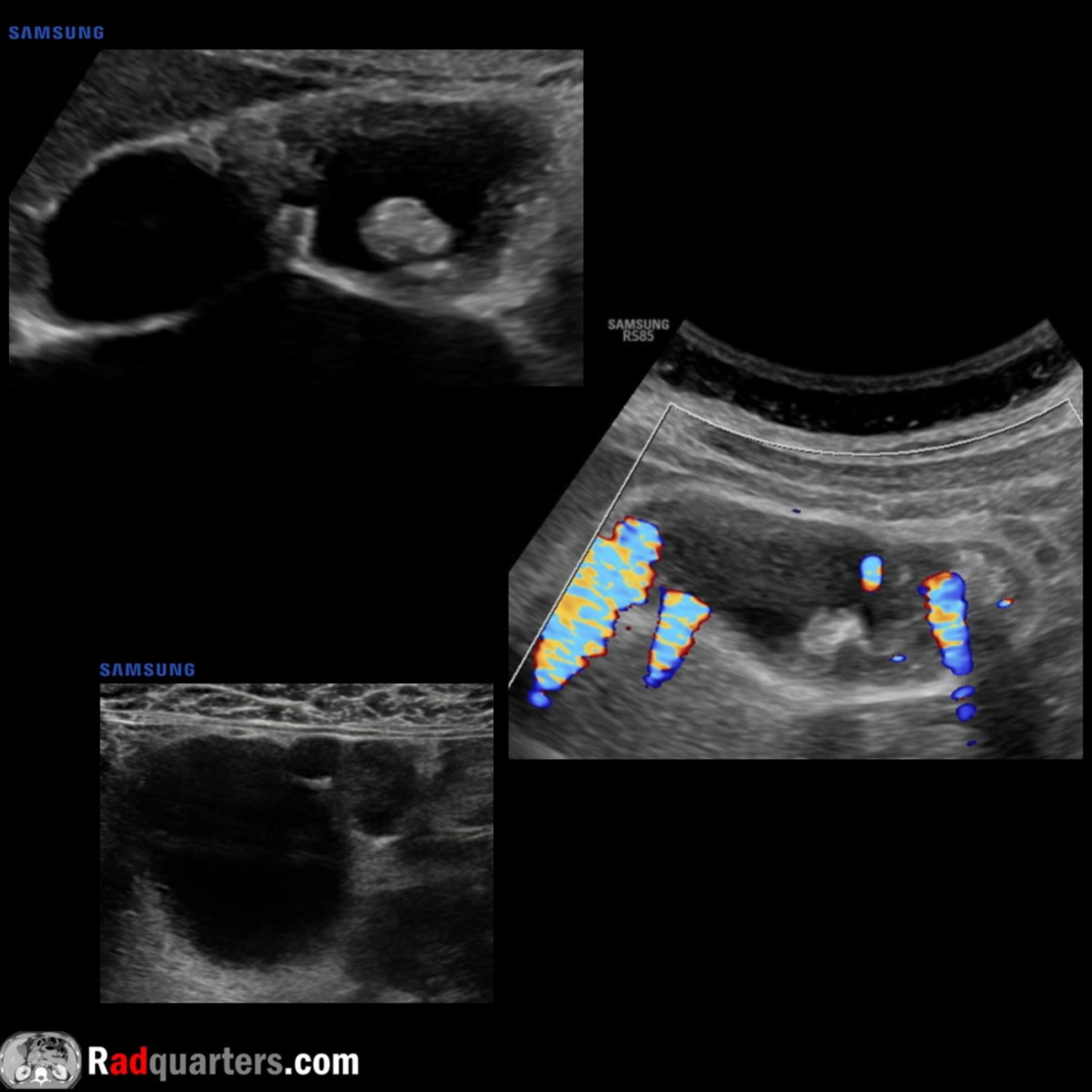
In this radiology lecture, we review the ultrasound appearance of adenomyomatosis of the gallbladder!Key teaching points include:Common cause of benign gallbladder wall thickening seen in up to 9% of patientsIncidence increases with ageUsually asymptomatic, but may be associated with sporadic RUQ painHyperplastic changes of gallbladder wall with mucosal overgrowth. Mucosal herniations protrude into muscular layer forming tiny, bile-filled cystic spaces = Rokitansky-Aschoff sinusesIf large, sinuses may appear as discrete cystic spaces in gallbladder wallCholesterol crystals in sinuses cause comet-tail reverberation artifact: Most common finding and highly specific for adenomyomatosis. Can exaggerate comet-tail with addition of color DopplerThree types: Focal/fundal, segmental/annular and diffuse. Regardless of type, comet-tail artifacts and/or cystic spaces are key to diagnosisFocal/fundal type: Most common. Often exhibits an “ovary on the gallbladder” appearance. Can be confused with a gallbladder mass. High-frequency linear transducer may be helpful to identify morphologySegmental/annular type: Narrows waist of gallbladder yielding a figure 8 or hourglass configuration. Gallstones and/or sludge often form in proximal lumen due to increased stasisIf necessary, MRI helpful for problem solving: T2 hyperintense pearl necklace/string of beads appearance sensitive and specificTo learn more about the Samsung RS85 Prestige ultrasound system, please visit: ⁠⁠⁠⁠⁠⁠⁠⁠⁠⁠https://www.bostonimaging.com/rs85-prestige-ultrasound-system-4⁠⁠⁠⁠⁠⁠⁠⁠⁠⁠Click the ⁠⁠⁠⁠⁠⁠⁠⁠⁠⁠YouTube Community tab⁠⁠⁠⁠⁠⁠⁠⁠⁠⁠ or follow on social media for bonus teaching material posted throughout the week!Website: ⁠⁠⁠⁠⁠⁠⁠⁠⁠⁠https://radquarters.com/⁠⁠⁠⁠⁠⁠⁠⁠⁠⁠Instagram: ⁠⁠⁠⁠⁠⁠⁠⁠⁠⁠https://www.instagram.com/radquarters/⁠⁠⁠⁠⁠⁠⁠⁠⁠⁠Facebook: ⁠⁠⁠⁠⁠⁠⁠⁠⁠⁠https://www.facebook.com/radquarters/⁠⁠⁠⁠⁠⁠⁠⁠⁠⁠X (Twitter): ⁠⁠⁠⁠⁠⁠⁠⁠⁠⁠https://twitter.com/radquarters⁠⁠⁠⁠⁠⁠⁠⁠⁠⁠Reddit: ⁠⁠⁠⁠⁠⁠⁠⁠⁠⁠⁠https://www.reddit.com/user/radiologistHQ/⁠⁠This video is for informational purposes only. It does not replace the advice or counsel of a doctor or healthcare professional.

Episode metadata supplied by the publisher feed · Published Oct 17, 2024

In this radiology lecture, we review the ultrasound appearance of adenomyomatosis of the gallbladder!Key teaching points include:Common cause of benign gallbladder wall thickening seen in up to 9% of patientsIncidence increases with ageUsually asymptomatic, but may be associated with sporadic RUQ painHyperplastic changes of gallbladder wall with mucosal overgrowth. Mucosal herniations protrude into muscular layer forming tiny, bile-filled cystic spaces = Rokitansky-Aschoff sinusesIf large, sinuses may appear as discrete cystic spaces in gallbladder wallCholesterol crystals in sinuses cause comet-tail reverberation artifact: Most common finding and highly specific for adenomyomatosis. Can exaggerate comet-tail with addition of color DopplerThree types: Focal/fundal, segmental/annular and diffuse. Regardless of type, comet-tail artifacts and/or cystic spaces are key to diagnosisFocal/fundal type: Most common. Often exhibits an “ovary on the gallbladder” appearance. Can be confused with a gallbladder mass. High-frequency linear transducer may be helpful to identify morphologySegmental/annular type: Narrows waist of gallbladder yielding a figure 8 or hourglass configuration. Gallstones and/or sludge often form in proximal lumen due to increased stasisIf necessary, MRI helpful for problem solving: T2 hyperintense pearl necklace/string of beads appearance sensitive and specificTo learn more about the Samsung RS85 Prestige ultrasound system, please visit: ⁠⁠⁠⁠⁠⁠⁠⁠⁠⁠https://www.bostonimaging.com/rs85-prestige-ultrasound-system-4⁠⁠⁠⁠⁠⁠⁠⁠⁠⁠Click the ⁠⁠⁠⁠⁠⁠⁠⁠⁠⁠YouTube Community tab⁠⁠⁠⁠⁠⁠⁠⁠⁠⁠ or follow on social media for bonus teaching material posted throughout the week!Website: ⁠⁠⁠⁠⁠⁠⁠⁠⁠⁠https://radquarters.com/⁠⁠⁠⁠⁠⁠⁠⁠⁠⁠Instagram: ⁠⁠⁠⁠⁠⁠⁠⁠⁠⁠https://www.instagram.com/radquarters/⁠⁠⁠⁠⁠⁠⁠⁠⁠⁠Facebook: ⁠⁠⁠⁠⁠⁠⁠⁠⁠⁠https://www.facebook.com/radquarters/⁠⁠⁠⁠⁠⁠⁠⁠⁠⁠X (Twitter): ⁠⁠⁠⁠⁠⁠⁠⁠⁠⁠https://twitter.com/radquarters⁠⁠⁠⁠⁠⁠⁠⁠⁠⁠Reddit: ⁠⁠⁠⁠⁠⁠⁠⁠⁠⁠⁠https://www.reddit.com/user/radiologistHQ/⁠⁠This video is for informational purposes only. It does not replace the advice or counsel of a doctor or healthcare professional.

PodParley-generated summary based on available episode metadata and transcript content.

NOW PLAYING

Ultrasound of Gallbladder Adenomyomatosis

0:00 8:02

No transcript for this episode yet

We transcribe on demand. Request one and we'll notify you when it's ready — usually under 10 minutes.

Frequently Asked Questions

How long is this episode of Radiology Lectures | Radquarters?

This episode is 8 minutes long.

When was this Radiology Lectures | Radquarters episode published?

This episode was published on October 17, 2024.

What is this episode about?

In this radiology lecture, we review the ultrasound appearance of adenomyomatosis of the gallbladder!Key teaching points include:Common cause of benign gallbladder wall thickening seen in up to 9% of patientsIncidence increases with ageUsually...

Can I download this Radiology Lectures | Radquarters episode?

Yes, you can download this episode by clicking the download button on the episode player, or subscribe to the podcast in your preferred podcast app for automatic downloads.
URL copied to clipboard!