
EPISODE · Dec 12, 2024 · 11 MIN
Ultrasound of Hydrosalpinx, Pyosalpinx & Tubo-ovarian Abscess
from Radiology Lectures | Radquarters · host Daniel J. Kowal, MD
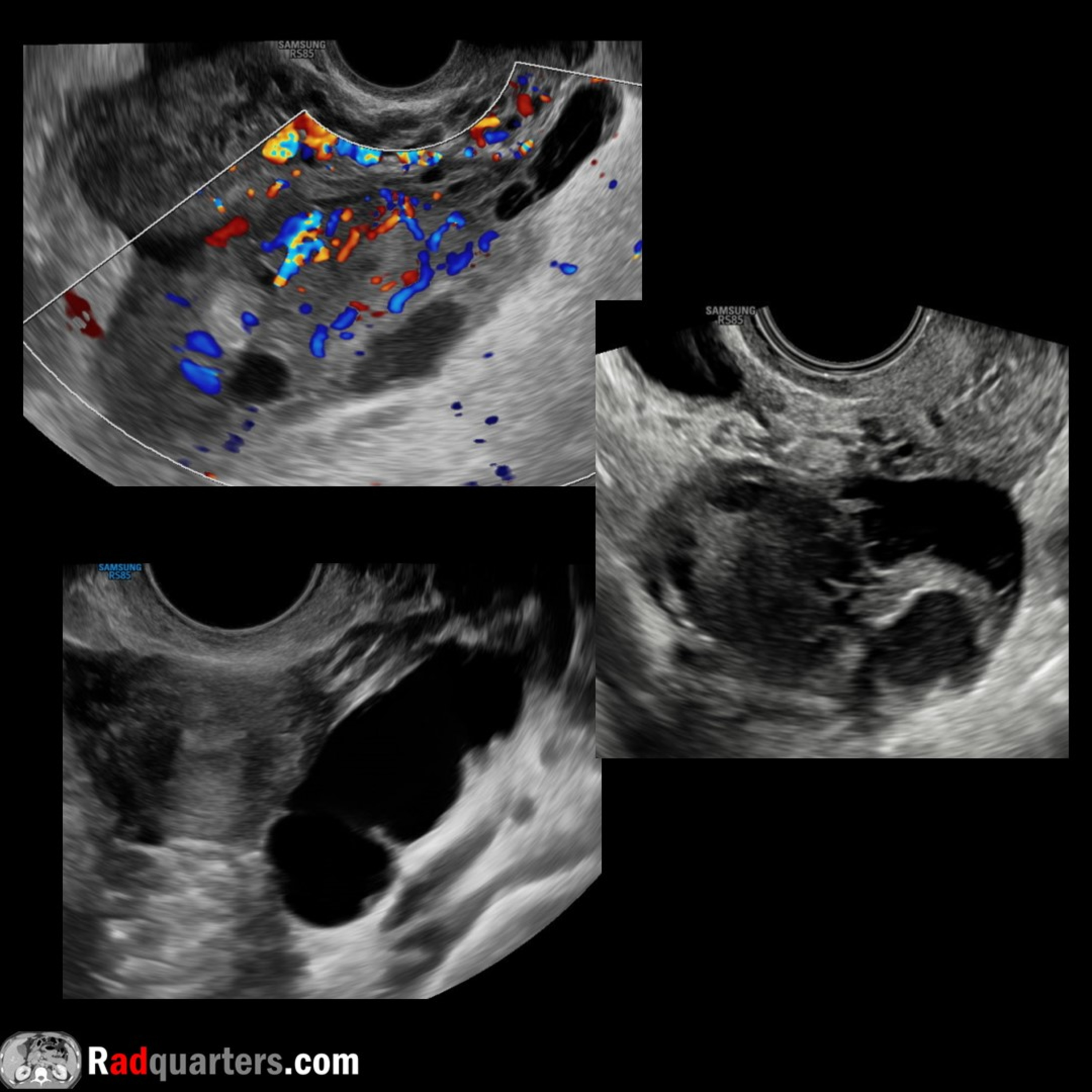
In this radiology lecture, we review the ultrasound appearance of hydrosalpinx, pyosalpinx and tubo-ovarian abscess!Key teaching points include:Hydrosalpinx = Fluid-filled, blocked fallopian tubeHydrosalpinx causes: Pelvic inflammatory disease (most common), endometriosis, prior surgery, adhesionsHydrosalpinx US: Thin-walled, tubular structure filled with anechoic simple fluid. Dilated tube may fold upon itself forming tubular C-shaped or S-shaped cystic mass. Incomplete septations commonWith chronic hydrosalpinx, may see “beads-on-a-string” sign: Short, round, 2-3 mm projections seen along inner tubal walls in cross section = Flattened, fibrotic remnants of endosalpingeal folds. Don’t confuse with solid mural nodulesO-RADS US v2022 management of hydrosalpinx = Imaging: None. Clinical: GynecologistPyosalpinx: Inflamed, blocked fallopian tube filled with purulent debris. Indicates pelvic inflammatory diseasePyosalpinx US: Thick-walled tubal structure filled with complex fluid. Like hydrosalpinx, typically conforms to a C or S-shape“Cogwheel” sign of pyosalpinx: Thickened endosalpingeal folds with surrounding tubal wall thickening. Typical of acute tubal inflammationTubal wall hyperemia more common with pyosalpinx than hydrosalpinxTubo-ovarian complex (TOC): With severe salpingo-oophoritis, ovary and tube adhere to each other. Can distinctly identify ovary from tube but cannot separate the two with transducer pressure. Tx: AntibioticsTubo-ovarian abscess (TOA): As pelvic inflammatory disease progresses, complete or near-complete loss of adnexal architecture with pockets of purulent fluid develop. Multiloculated mass with septations, irregular margins, may be bilateral. Tx: Antibiotics, percutaneous drainage, surgeryTo learn more about the Samsung RS85 Prestige ultrasound system, please visit: https://www.bostonimaging.com/rs85-prestige-ultrasound-system-4Click the YouTube Community tab or follow on social media for bonus teaching material posted throughout the week!Website: https://radquarters.com/Instagram: https://www.instagram.com/radquarters/Facebook: https://www.facebook.com/radquarters/X (Twitter): https://twitter.com/radquartersReddit: https://www.reddit.com/user/radiologistHQ/This video is for informational purposes only. It does not replace the advice or counsel of a doctor or healthcare professional.
Embed this episode
NOW PLAYING
Ultrasound of Hydrosalpinx, Pyosalpinx & Tubo-ovarian Abscess
No transcript for this episode yet
Similar Episodes
No similar episodes found.
Similar Podcasts
No similar podcasts found.