Ultrasound of Intussusception episode artwork

EPISODE · May 25, 2023 · 8 MIN

Ultrasound of Intussusception

from Radiology Lectures | Radquarters · host Daniel J. Kowal, MD

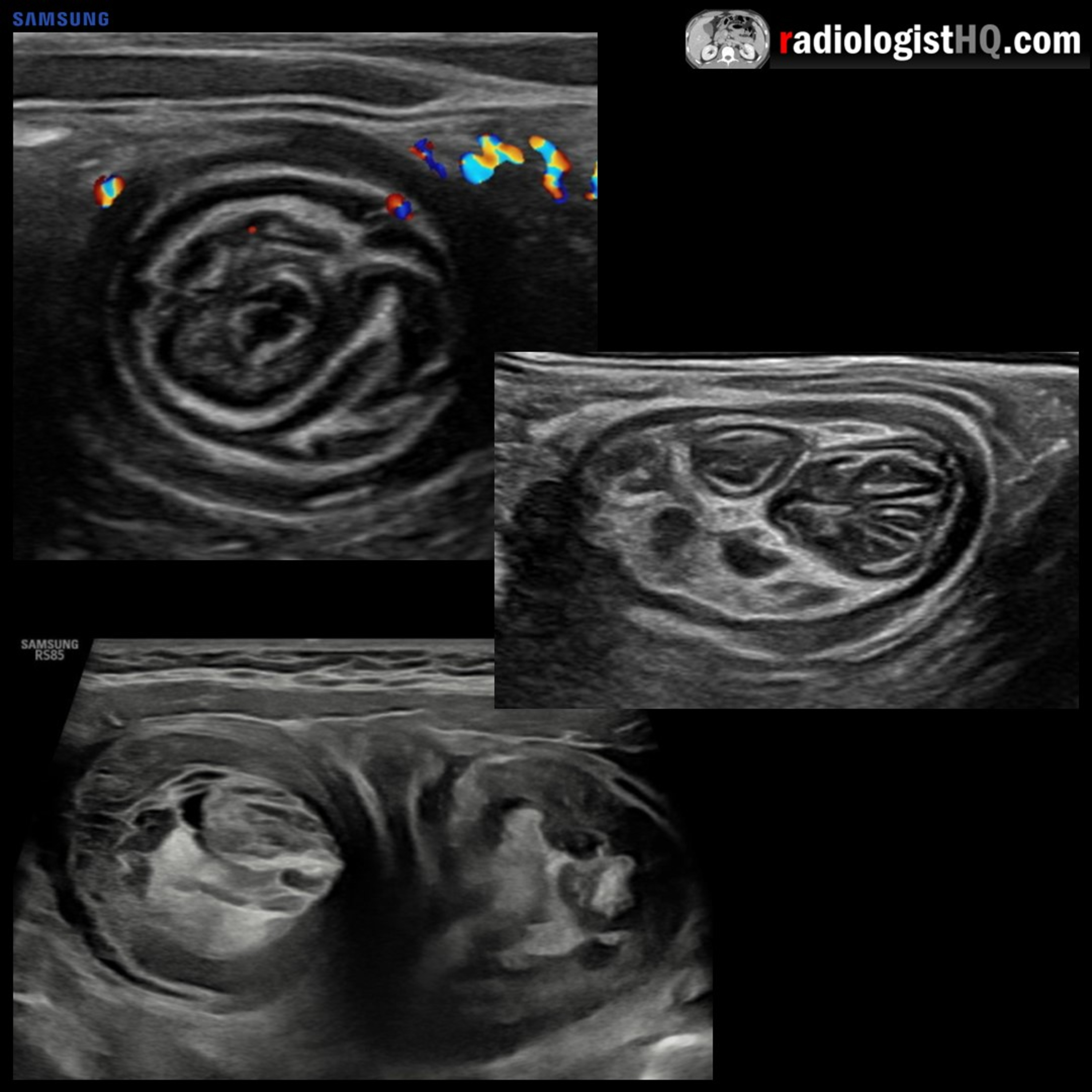
In this radiology lecture, we review the ultrasound appearance of ileocolic and small bowel-small bowel intussusception in children! Key teaching points include: Intussusception occurs when bowel is pulled into itself or into neighboring bowel. Intussusceptum is the prolapsing bowel pulled into intussuscipiens which receives the bowel. Two major types: Ileocolic and small bowel-small bowel. If ileocolic not reduced = Bowel ischemia and perforation. Most occur in children beyond 3 months of age. Usually no lead point in children (unlike adults), suspected that due to hypertrophic lymphoid tissue after infection. Clinical triad of colicky abdominal pain, vomiting, palpable abdominal mass seen in less than 50% of cases. Red-currant jelly stool = Stool mixed with blood and mucus, can be seen with bowel ischemia. Ultrasound gold standard in diagnosis: Sensitivity and specificity 98%, false negative rate less than 1%. “Target” sign (short axis) and “pseudokidney” sign (long axis) may be seen. Findings suggesting ileocolic (as opposed to small bowel-small bowel) intussusception: Location in right lower quadrant with absent normal ileocolic junction, hyperechoic center indicating mesenteric fat, diameter of hyperechoic core greater than outer wall, lymph nodes inside intussusception, larger AP diameter greater than 2 cm, and longer length greater than 3 cm. Treatment of ileocolic intussusception: Enema with air or contrast material. Findings suspicious for ischemia/necrosis and increased risk of enema reduction failure: Fluid trapped within the intussuscipiens, lack of internal vascular flow on Doppler within the intussusceptum, and irregular bowel wall or decreased bowel wall vascularity. To learn more about the Samsung RS85 Prestige ultrasound system, please visit: ⁠⁠https://www.bostonimaging.com/rs85-prestige-ultrasound-system-4⁠⁠ Click the ⁠⁠YouTube Community tab⁠⁠ or follow on social media for bonus teaching material posted throughout the week! Website: ⁠⁠https://radiologisthq.com/⁠⁠ Instagram: ⁠⁠https://www.instagram.com/radiologistHQ/⁠⁠ Facebook: ⁠⁠https://www.facebook.com/radiologistHeadQuarters/⁠⁠ Twitter: ⁠⁠https://twitter.com/radiologistHQ⁠⁠ Reddit: ⁠⁠https://www.reddit.com/user/radiologistHQ/

In this radiology lecture, we review the ultrasound appearance of ileocolic and small bowel-small bowel intussusception in children! Key teaching points include: Intussusception occurs when bowel is pulled into itself or into neighboring bowel. Intussusceptum is the prolapsing bowel pulled into intussuscipiens which receives the bowel. Two major types: Ileocolic and small bowel-small bowel. If ileocolic not reduced = Bowel ischemia and perforation. Most occur in children beyond 3 months of age. Usually no lead point in children (unlike adults), suspected that due to hypertrophic lymphoid tissue after infection. Clinical triad of colicky abdominal pain, vomiting, palpable abdominal mass seen in less than 50% of cases. Red-currant jelly stool = Stool mixed with blood and mucus, can be seen with bowel ischemia. Ultrasound gold standard in diagnosis: Sensitivity and specificity 98%, false negative rate less than 1%. “Target” sign (short axis) and “pseudokidney” sign (long axis) may be seen. Findings suggesting ileocolic (as opposed to small bowel-small bowel) intussusception: Location in right lower quadrant with absent normal ileocolic junction, hyperechoic center indicating mesenteric fat, diameter of hyperechoic core greater than outer wall, lymph nodes inside intussusception, larger AP diameter greater than 2 cm, and longer length greater than 3 cm. Treatment of ileocolic intussusception: Enema with air or contrast material. Findings suspicious for ischemia/necrosis and increased risk of enema reduction failure: Fluid trapped within the intussuscipiens, lack of internal vascular flow on Doppler within the intussusceptum, and irregular bowel wall or decreased bowel wall vascularity. To learn more about the Samsung RS85 Prestige ultrasound system, please visit: ⁠⁠https://www.bostonimaging.com/rs85-prestige-ultrasound-system-4⁠⁠ Click the ⁠⁠YouTube Community tab⁠⁠ or follow on social media for bonus teaching material posted throughout the week! Website: ⁠⁠https://radiologisthq.com/⁠⁠ Instagram: ⁠⁠https://www.instagram.com/radiologistHQ/⁠⁠ Facebook: ⁠⁠https://www.facebook.com/radiologistHeadQuarters/⁠⁠ Twitter: ⁠⁠https://twitter.com/radiologistHQ⁠⁠ Reddit: ⁠⁠https://www.reddit.com/user/radiologistHQ/

NOW PLAYING

Ultrasound of Intussusception

0:00 8:39

No transcript for this episode yet

We transcribe on demand. Request one and we'll notify you when it's ready — usually under 10 minutes.

Frequently Asked Questions

How long is this episode of Radiology Lectures | Radquarters?

This episode is 8 minutes long.

When was this Radiology Lectures | Radquarters episode published?

This episode was published on May 25, 2023.

What is this episode about?

In this radiology lecture, we review the ultrasound appearance of ileocolic and small bowel-small bowel intussusception in children! Key teaching points include: Intussusception occurs when bowel is pulled into itself or into neighboring bowel. ...

Can I download this Radiology Lectures | Radquarters episode?

Yes, you can download this episode by clicking the download button on the episode player, or subscribe to the podcast in your preferred podcast app for automatic downloads.
URL copied to clipboard!