Ultrasound of Polycystic Ovarian Syndrome episode artwork

EPISODE · Apr 27, 2023 · 6 MIN

Ultrasound of Polycystic Ovarian Syndrome

from Radiology Lectures | Radquarters · host Daniel J. Kowal, MD

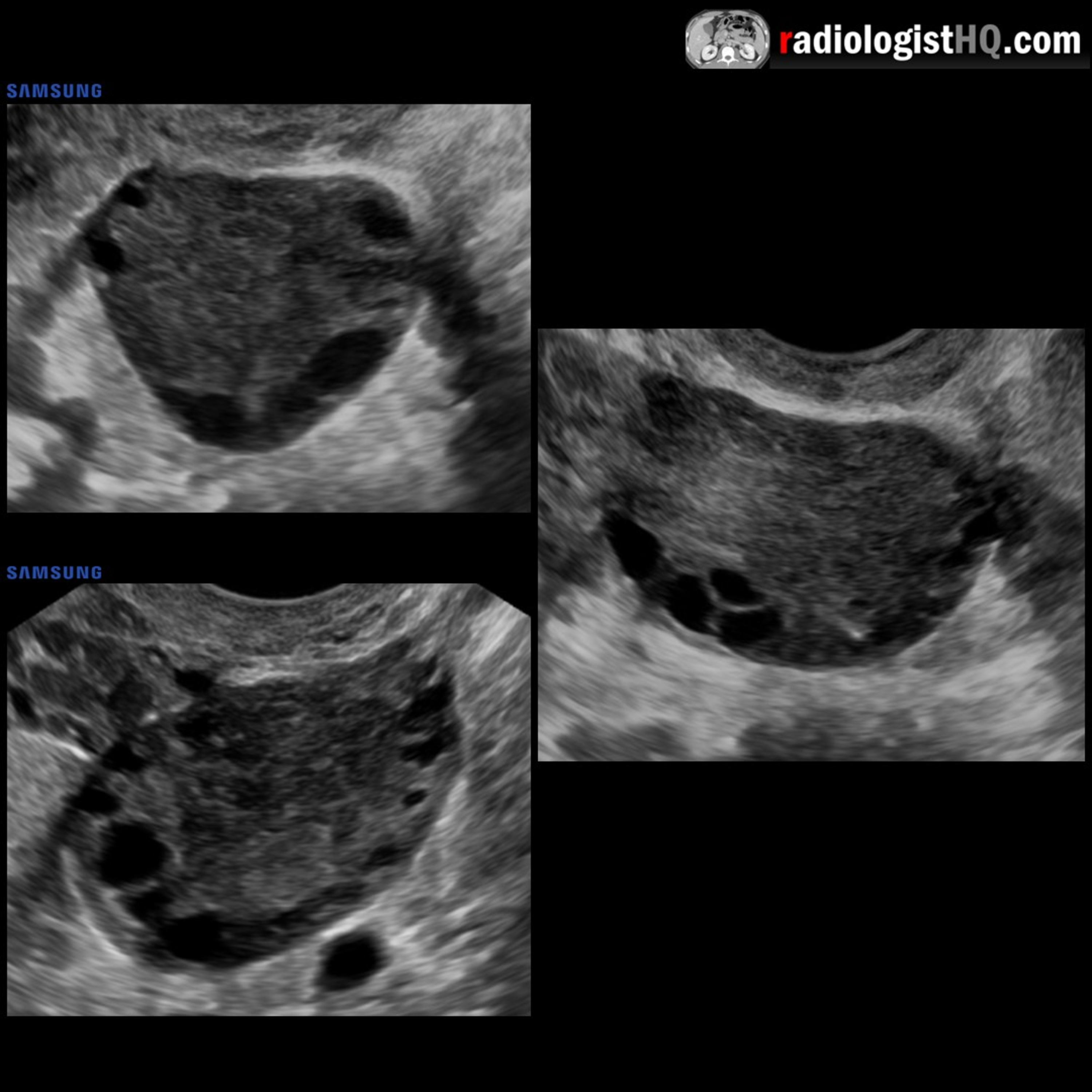
In this radiology lecture, we review the ultrasound appearance of polycystic ovarian syndrome (PCOS)! Key teaching points include: PCOS often presents with the clinical triad of oligomenorrhea and/or anovulation, hirsutism, and obesity. Associated with subfertility and recurrent pregnancy loss. Rotterdam criteria (2003) states that PCOS diagnosis requires at least two of the following: Oligo- or anovulation (ovulatory dysfunction), hyperandrogenism (clinical and/or biochemical signs), and polycystic ovarian morphology on ultrasound. Ovaries can be sonographically normal in PCOS. “Hyperandrogenic anovulation” proposed as a more accurate term. Ovaries can also appear polycystic on ultrasound without clinical diagnosis of PCOS. Rotterdam description of polycystic ovaries: 12 or more follicles 2-9 mm in size, and/or ovarian volume greater than 10 cc in at least one ovary (with no dominant cysts). Specific diagnostic cutoffs debated, and 20-25 or more follicles has been more recently suggested as a more accurate cutoff. Supportive morphologic features of PCOS include the “string of pearls sign” (peripheral location of follicles) and prominent, hyperechoic central ovarian stroma. Ovarian morphology typically more important than ovarian size, although a single enlarged, polycystic ovary sufficiently meets ultrasound criteria for PCOS. The term “polycystic” is generally incorrect and “multifollicular” has been offered as a more accurate ultrasound description, but PCOS remains the most widely used term. In post-menopausal women with new or worsening hyperandrogenism, also consider androgen-secreting tumors of ovaries or adrenal glands. To learn more about the Samsung RS85 Prestige ultrasound system, please visit: ⁠https://www.bostonimaging.com/rs85-prestige-ultrasound-system-4⁠ Click the ⁠YouTube Community tab⁠ or follow on social media for bonus teaching material posted throughout the week! Website: ⁠https://radiologisthq.com/⁠ Instagram: ⁠https://www.instagram.com/radiologistHQ/⁠ Facebook: ⁠https://www.facebook.com/radiologistHeadQuarters/⁠ Twitter: ⁠https://twitter.com/radiologistHQ⁠ Reddit: ⁠https://www.reddit.com/user/radiologistHQ/

Episode metadata supplied by the publisher feed · Published Apr 27, 2023

In this radiology lecture, we review the ultrasound appearance of polycystic ovarian syndrome (PCOS)! Key teaching points include: PCOS often presents with the clinical triad of oligomenorrhea and/or anovulation, hirsutism, and obesity. Associated with subfertility and recurrent pregnancy loss. Rotterdam criteria (2003) states that PCOS diagnosis requires at least two of the following: Oligo- or anovulation (ovulatory dysfunction), hyperandrogenism (clinical and/or biochemical signs), and polycystic ovarian morphology on ultrasound. Ovaries can be sonographically normal in PCOS. “Hyperandrogenic anovulation” proposed as a more accurate term. Ovaries can also appear polycystic on ultrasound without clinical diagnosis of PCOS. Rotterdam description of polycystic ovaries: 12 or more follicles 2-9 mm in size, and/or ovarian volume greater than 10 cc in at least one ovary (with no dominant cysts). Specific diagnostic cutoffs debated, and 20-25 or more follicles has been more recently suggested as a more accurate cutoff. Supportive morphologic features of PCOS include the “string of pearls sign” (peripheral location of follicles) and prominent, hyperechoic central ovarian stroma. Ovarian morphology typically more important than ovarian size, although a single enlarged, polycystic ovary sufficiently meets ultrasound criteria for PCOS. The term “polycystic” is generally incorrect and “multifollicular” has been offered as a more accurate ultrasound description, but PCOS remains the most widely used term. In post-menopausal women with new or worsening hyperandrogenism, also consider androgen-secreting tumors of ovaries or adrenal glands. To learn more about the Samsung RS85 Prestige ultrasound system, please visit: ⁠https://www.bostonimaging.com/rs85-prestige-ultrasound-system-4⁠ Click the ⁠YouTube Community tab⁠ or follow on social media for bonus teaching material posted throughout the week! Website: ⁠https://radiologisthq.com/⁠ Instagram: ⁠https://www.instagram.com/radiologistHQ/⁠ Facebook: ⁠https://www.facebook.com/radiologistHeadQuarters/⁠ Twitter: ⁠https://twitter.com/radiologistHQ⁠ Reddit: ⁠https://www.reddit.com/user/radiologistHQ/

PodParley-generated summary based on available episode metadata and transcript content.

NOW PLAYING

Ultrasound of Polycystic Ovarian Syndrome

0:00 6:54

No transcript for this episode yet

We transcribe on demand. Request one and we'll notify you when it's ready — usually under 10 minutes.

Frequently Asked Questions

How long is this episode of Radiology Lectures | Radquarters?

This episode is 6 minutes long.

When was this Radiology Lectures | Radquarters episode published?

This episode was published on April 27, 2023.

What is this episode about?

In this radiology lecture, we review the ultrasound appearance of polycystic ovarian syndrome (PCOS)! Key teaching points include: PCOS often presents with the clinical triad of oligomenorrhea and/or anovulation, hirsutism, and obesity. Associated...

Can I download this Radiology Lectures | Radquarters episode?

Yes, you can download this episode by clicking the download button on the episode player, or subscribe to the podcast in your preferred podcast app for automatic downloads.
URL copied to clipboard!