Ultrasound of Trigger Finger (Stenosing Tenosynovitis) episode artwork

EPISODE · Jan 16, 2025 · 11 MIN

Ultrasound of Trigger Finger (Stenosing Tenosynovitis)

from Radiology Lectures | Radquarters · host Daniel J. Kowal, MD

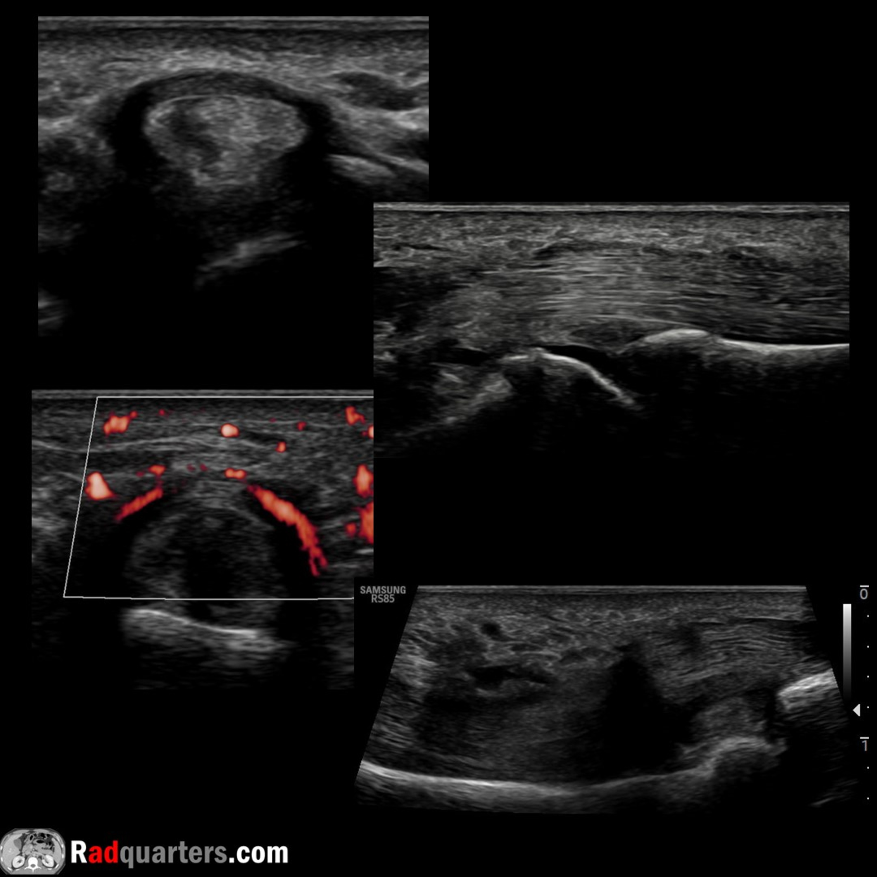
In this radiology lecture, we review the ultrasound appearance of trigger finger!Key teaching points include:Pulleys are fibrous retinacula on ventral finger that secure flexor tendons to phalanges preventing tendon displacement and bowstringing with finger flexionFinger has 5 annular pulleys. Odd-numbered at joints, even-numbered at phalanges: A1, A3, A5 are located about the MCP, PIP, DIP joints, respectively. A2 is located at the proximal phalanx, and A4 at the middle phalanxCruciform pulleys lie between annular pulleys, but are not usually well-seen on ultrasoundTrigger finger, also known as stenosing tenosynovitis, is characterized by impaired flexor tendon movement due to thickened pulley leading to tendon constrictionMost common at A1 pulley, but can also occur at A2/A3 pulleys, palmar aponeurosis (A0) and wristSymptoms: Triggering/locking when flexed, painful snapping when extended, pain, joint stiffnessMost common in female patients, history of diabetes mellitus, and rheumatoid arthritisOften idiopathic, can occur with repetitive microinjury (flexion-extension). Can also be post-traumatic or due to compressive mass/cystTx: Splinting, NSAIDs, US-guided corticosteroid injection, surgical releaseA1 pulley thickness cutoff = 0.62 mm*. Mean normal thickness = 0.5 mm, range with trigger finger 1.1-2.9 mm**. Comparison with asymptomatic side helpfulAdditional findings: Pulley hyperemia, nodular tendon thickening (tendinosis) or tear, buckling of flexor tendon on dynamic imaging, “dark tendon” sign (anisotropic hypoechogenicity due to tendon constriction), synovial sheath effusion (acute), and peri-pulley cyst/cystic degenerationReferences:*Spirig A, Juon B, Banz Y, Rieben R, Vogelin E. Correlation between sonographic and in vivo measurement of A1 pulleys in trigger fingers. Ultrasound Med Biol 2016; 42:1482–1490.**Guerini H, Pessis E, Theumann N, Le Quintrec JS, Campagna R, Chevrot A, Feydy A, Drapé JL. Sonographic appearance of trigger fingers. J Ultrasound Med. 2008 Oct;27(10):1407-13.Bianchi S, Gitto S, Draghi F. Ultrasound Features of Trigger Finger: Review of the Literature. J Ultrasound Med. 2019 Dec;38(12):3141-3154.Shohda E, Sheta RA. Misconceptions about trigger finger: a scoping review. Definition, pathophysiology, site of lesion, etiology. Trigger finger solving a maze. Adv Rheumatol. 2024 Jul 11;64(1):53.To learn more about the Samsung RS85 Prestige ultrasound system, please visit: ⁠⁠⁠⁠⁠⁠⁠⁠⁠⁠⁠⁠https://www.bostonimaging.com/rs85-prestige-ultrasound-system-4⁠⁠⁠⁠⁠⁠⁠⁠⁠⁠⁠⁠⁠Click the ⁠⁠⁠⁠⁠⁠⁠⁠⁠⁠⁠⁠⁠YouTube Community tab⁠⁠⁠⁠⁠⁠⁠⁠⁠⁠⁠⁠⁠ or follow on social media for bonus teaching material posted throughout the week!Website: ⁠⁠⁠⁠⁠⁠⁠⁠⁠⁠⁠⁠⁠https://radquarters.com/⁠⁠⁠⁠⁠⁠⁠⁠⁠⁠⁠⁠⁠Instagram: ⁠⁠⁠⁠⁠⁠⁠⁠⁠⁠⁠⁠⁠https://www.instagram.com/radquarters/⁠⁠⁠⁠⁠⁠⁠⁠⁠⁠⁠⁠⁠Facebook: ⁠⁠⁠⁠⁠⁠⁠⁠⁠⁠⁠⁠⁠https://www.facebook.com/radquarters/⁠⁠⁠⁠⁠⁠⁠⁠⁠⁠⁠⁠⁠X (Twitter): ⁠⁠⁠⁠⁠⁠⁠⁠⁠⁠⁠⁠⁠https://twitter.com/radquarters⁠⁠⁠⁠⁠⁠⁠⁠⁠⁠⁠⁠⁠Reddit: ⁠⁠⁠⁠⁠⁠⁠⁠⁠⁠⁠⁠⁠⁠https://www.reddit.com/user/radiologistHQ/⁠⁠This video is for informational purposes only. It does not replace the advice or counsel of a doctor or healthcare professional.

In this radiology lecture, we review the ultrasound appearance of trigger finger!Key teaching points include:Pulleys are fibrous retinacula on ventral finger that secure flexor tendons to phalanges preventing tendon displacement and bowstringing with finger flexionFinger has 5 annular pulleys. Odd-numbered at joints, even-numbered at phalanges: A1, A3, A5 are located about the MCP, PIP, DIP joints, respectively. A2 is located at the proximal phalanx, and A4 at the middle phalanxCruciform pulleys lie between annular pulleys, but are not usually well-seen on ultrasoundTrigger finger, also known as stenosing tenosynovitis, is characterized by impaired flexor tendon movement due to thickened pulley leading to tendon constrictionMost common at A1 pulley, but can also occur at A2/A3 pulleys, palmar aponeurosis (A0) and wristSymptoms: Triggering/locking when flexed, painful snapping when extended, pain, joint stiffnessMost common in female patients, history of diabetes mellitus, and rheumatoid arthritisOften idiopathic, can occur with repetitive microinjury (flexion-extension). Can also be post-traumatic or due to compressive mass/cystTx: Splinting, NSAIDs, US-guided corticosteroid injection, surgical releaseA1 pulley thickness cutoff = 0.62 mm*. Mean normal thickness = 0.5 mm, range with trigger finger 1.1-2.9 mm**. Comparison with asymptomatic side helpfulAdditional findings: Pulley hyperemia, nodular tendon thickening (tendinosis) or tear, buckling of flexor tendon on dynamic imaging, “dark tendon” sign (anisotropic hypoechogenicity due to tendon constriction), synovial sheath effusion (acute), and peri-pulley cyst/cystic degenerationReferences:*Spirig A, Juon B, Banz Y, Rieben R, Vogelin E. Correlation between sonographic and in vivo measurement of A1 pulleys in trigger fingers. Ultrasound Med Biol 2016; 42:1482–1490.**Guerini H, Pessis E, Theumann N, Le Quintrec JS, Campagna R, Chevrot A, Feydy A, Drapé JL. Sonographic appearance of trigger fingers. J Ultrasound Med. 2008 Oct;27(10):1407-13.Bianchi S, Gitto S, Draghi F. Ultrasound Features of Trigger Finger: Review of the Literature. J Ultrasound Med. 2019 Dec;38(12):3141-3154.Shohda E, Sheta RA. Misconceptions about trigger finger: a scoping review. Definition, pathophysiology, site of lesion, etiology. Trigger finger solving a maze. Adv Rheumatol. 2024 Jul 11;64(1):53.To learn more about the Samsung RS85 Prestige ultrasound system, please visit: ⁠⁠⁠⁠⁠⁠⁠⁠⁠⁠⁠⁠https://www.bostonimaging.com/rs85-prestige-ultrasound-system-4⁠⁠⁠⁠⁠⁠⁠⁠⁠⁠⁠⁠⁠Click the ⁠⁠⁠⁠⁠⁠⁠⁠⁠⁠⁠⁠⁠YouTube Community tab⁠⁠⁠⁠⁠⁠⁠⁠⁠⁠⁠⁠⁠ or follow on social media for bonus teaching material posted throughout the week!Website: ⁠⁠⁠⁠⁠⁠⁠⁠⁠⁠⁠⁠⁠https://radquarters.com/⁠⁠⁠⁠⁠⁠⁠⁠⁠⁠⁠⁠⁠Instagram: ⁠⁠⁠⁠⁠⁠⁠⁠⁠⁠⁠⁠⁠https://www.instagram.com/radquarters/⁠⁠⁠⁠⁠⁠⁠⁠⁠⁠⁠⁠⁠Facebook: ⁠⁠⁠⁠⁠⁠⁠⁠⁠⁠⁠⁠⁠https://www.facebook.com/radquarters/⁠⁠⁠⁠⁠⁠⁠⁠⁠⁠⁠⁠⁠X (Twitter): ⁠⁠⁠⁠⁠⁠⁠⁠⁠⁠⁠⁠⁠https://twitter.com/radquarters⁠⁠⁠⁠⁠⁠⁠⁠⁠⁠⁠⁠⁠Reddit: ⁠⁠⁠⁠⁠⁠⁠⁠⁠⁠⁠⁠⁠⁠https://www.reddit.com/user/radiologistHQ/⁠⁠This video is for informational purposes only. It does not replace the advice or counsel of a doctor or healthcare professional.

NOW PLAYING

Ultrasound of Trigger Finger (Stenosing Tenosynovitis)

0:00 11:32

No transcript for this episode yet

We transcribe on demand. Request one and we'll notify you when it's ready — usually under 10 minutes.

Frequently Asked Questions

How long is this episode of Radiology Lectures | Radquarters?

This episode is 11 minutes long.

When was this Radiology Lectures | Radquarters episode published?

This episode was published on January 16, 2025.

What is this episode about?

In this radiology lecture, we review the ultrasound appearance of trigger finger!Key teaching points include:Pulleys are fibrous retinacula on ventral finger that secure flexor tendons to phalanges preventing tendon displacement and bowstringing...

Can I download this Radiology Lectures | Radquarters episode?

Yes, you can download this episode by clicking the download button on the episode player, or subscribe to the podcast in your preferred podcast app for automatic downloads.
URL copied to clipboard!